This site uses cookies to improve your experience. To help us insure we adhere to various privacy regulations, please select your country/region of residence. If you do not select a country, we will assume you are from the United States. Select your Cookie Settings or view our Privacy Policy and Terms of Use.
Cookie Settings
Cookies and similar technologies are used on this website for proper function of the website, for tracking performance analytics and for marketing purposes. We and some of our third-party providers may use cookie data for various purposes. Please review the cookie settings below and choose your preference.
Used for the proper function of the website
Used for monitoring website traffic and interactions
Cookie Settings
Cookies and similar technologies are used on this website for proper function of the website, for tracking performance analytics and for marketing purposes. We and some of our third-party providers may use cookie data for various purposes. Please review the cookie settings below and choose your preference.
Strictly Necessary: Used for the proper function of the website
Performance/Analytics: Used for monitoring website traffic and interactions
Echocardiogram, CT aortogram and late gadolinium imaging of the aorta have been shown in figure 1. The coronaryangiogram was normal. Figure 1 (A) Two-dimensional echocardiogram short-axis basal view showing aortic valve; (B) volume-rendered CT aortogram. The renal and carotid Doppler tests were normal.
Finally, do a coronaryangiogram Possible alternative to pacing is to give a beta-1 agonist to increase heart rate. EKG with paced complexes shown below shows much narrower QRS complex and echocardiogram showed improved LV systolic function primarily due to improvement in LV dyssynchrony. (J J Am Coll Cardiol.
Workup including routine laboratory results, 12-lead electrocardiogram (ECG), echocardiogram, and coronaryangiogram was non-specific. During the intravenous lacosamide infusion, the patient developed sudden cardiac arrest caused by ventricular arrhythmias necessitating resuscitation.
And then a slightly more remote past ECG Old inferior MI The patient's previous echocardiogram report was viewed: Decreased LV systolic performance, estimated left ventricular ejection fraction is 35%. Case continued The patient underwent an emergency formal echocardiogram and it was unchanged. Cath Lab activation was cancelled.
The routine laboratory results, imaging study, coronaryangiogram, and echocardiogram (ECG) were normal. Case We report the case of a 20-year-old man with variants in SCN5A and RyR2 genes who was resuscitated from sudden cardiac death during sleep due to a ventricular fibrillation. The patient did not have underlying diseases.
Her ejection fraction was 66% ejection fraction with a fistula between the right sinus of Valsalva and the right atrium on transthoracic echocardiogram (TTE) which was also seen on transesophageal echocardiogram (TEE). Her heart failure was due to the fistula as she had no coronary artery disease on coronaryangiogram.
Transthoracic echocardiogram (TTE) showed an ejection fraction (EF) of 40% and a moderate-large pericardial effusion with signs of tamponade. Intra-operative transesophageal echocardiogram (TEE) post-decannulation showed a normal EF without segmental abnormalities. A repeat coronaryangiogram was unremarkable.
Echocardiogram is indicated (Correct) C. Start aspirin and Plavix Correct answer: (B) (B) Echocardiogram is indicated. Which of the following is the best statement to describe further clinical management? No further workup is indicated B. Start furosemide for diuresis D. Start with a Free Trial.
It’s judicious, then, to arrange for coronaryangiogram. Coronary occlusion, however, might be present concurrently with subendocardial ischemia on the time-zero ECG, or evolve into such. Proximal LAD disease with/without a) and b) It seemed quite apparent that this was an Acute Coronary Syndrome. CoronaryAngiogram 1.
He visited an outpatient clinic for it and an echocardiogram and exercise stress test was normal. In the meantime, cardiology consultant sees the patient and performs a bedside echocardiogram which revealed no major wall motion abnormalities. Here is the coronaryangiogram: A distal thrombotic right coronary artery (RCA) occlusion !
Angiogram showed a culprit lesion of 100% stenosis to the right coronary artery and 100% stenosis of the right posterior descending artery, both with TIMI 0 flow. Echocardiogram the following day showed a left ventricular ejection fraction of 52% (+/- 5%) with hypokinesis of the basal-mid inferior and inferoseptal myocardium.
Indeed, bedside Echocardiogram revealed severe left ventricular impairment of Takotsubo cardiomyopathy. The coronaryangiogram revealed no critical stenosis, or acute plaque ulceration. Furthermore, pertinent electrolyte values (e.g. potassium) were within normal parameter.
See this case: what do you think the echocardiogram shows in this case? We investigated the incidence of an acutely occluded coronary in patients presenting with STE-aVR with multi-lead ST depression. All electrocardiograms (ECGs) and coronaryangiograms were blindly analyzed by experienced cardiologists.
The diagnostic coronaryangiogram identified only minimal coronary artery disease, but there was a severely calcified, ‘immobile’ aortic valve. Aortic angiogram did not reveal aortic dissection. During the procedure, the patient had an increasing oxygen requirement and was intubated for airway protection and oxygenation.
More troponin values were measured at the cardiac center: 2327- 267 ng/L 0821- 355 ng/L 1108- 305 ng/L An echocardiogram on day three of the patients admission showed an ejection fraction of 46% with abnormal basal inferior and basal lateral segments, and severe aortic stenosis.
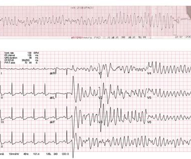
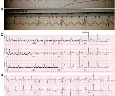
CT coronaryangiogram showed a hypoplastic RCA and dominant LCx. It is reasonable to perform an echocardiogram to evaluate LV function. Figure-5: Long lead II recording on oral flecainide ( 10 minutes of continuous recording each line being 1-minute long ). No PVCs are seen. There were no plaques or stenoses.
We organize all of the trending information in your field so you don't have to. Join thousands of users and stay up to date on the latest articles your peers are reading.
You know about us, now we want to get to know you!
Let's personalize your content
Let's get even more personalized
We recognize your account from another site in our network, please click 'Send Email' below to continue with verifying your account and setting a password.


Let's personalize your content