This site uses cookies to improve your experience. To help us insure we adhere to various privacy regulations, please select your country/region of residence. If you do not select a country, we will assume you are from the United States. Select your Cookie Settings or view our Privacy Policy and Terms of Use.
Cookie Settings
Cookies and similar technologies are used on this website for proper function of the website, for tracking performance analytics and for marketing purposes. We and some of our third-party providers may use cookie data for various purposes. Please review the cookie settings below and choose your preference.
Used for the proper function of the website
Used for monitoring website traffic and interactions
Cookie Settings
Cookies and similar technologies are used on this website for proper function of the website, for tracking performance analytics and for marketing purposes. We and some of our third-party providers may use cookie data for various purposes. Please review the cookie settings below and choose your preference.
Strictly Necessary: Used for the proper function of the website
Performance/Analytics: Used for monitoring website traffic and interactions
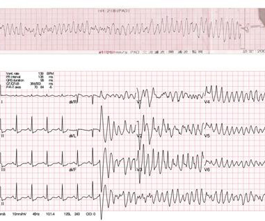
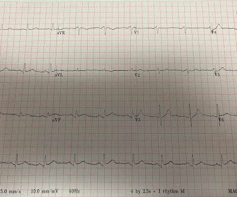
Post cath ECG: Now there are hyperacute T-waves again, and recurrent ST depression in V2 This ECG would normally diagnostic of OMI until proven otherwise No further troponins were measured, but it looks like there is recurrent OMI Next day: A CT CoronaryAngiogram was done (CTCA) CARDIAC MORPHOLOGY AND FUNCTION: 1. IMPRESSION: 1.
CT coronaryangiograms are increasing in popularity as a non-invasive screening test for detecting blocks in coronary arteries. Coronary arteries are blood vessels supplying oxygenated blood to the heart. Angiograms are images of blood vessels, usually obtained by injecting medications for contrast from body structures.
Sleep & Heart Disease Cardiac CT - CAC Scores & CT CoronaryAngiograms. Understanding Blood Pressure. The Role of Inflammation. Formulas For Estimating Benefit & Risk. The Reducing Risk module is where we put it all together.
A CTCA provides much more anatomical detail and can identify advanced plaque often missed by CT Coronary Artery Calcium Score scans alone. CT Coronary Artery Calcium Score Scan CT Coronary Artery Calcium Score CT CoronaryAngiogram As you can see from the above images, the CTCA provides far more anatomical detail.
Sleep & Heart Disease Cardiac CT - CAC Scores & CT CoronaryAngiograms. Understanding Blood Pressure. The Role of Inflammation. Formulas For Estimating Benefit & Risk. I'm Interested The Reducing Risk module is where we put it all together.
A CT Coronaryangiogram was ordered. Here are the results: --Minimally obstructive coronary artery disease. --LAD Although a lesion is not visible anatomically on this CT scan, coronary catheter angiography could be considered based on Cardiology evaluation." A repeat troponin returned at 0.45 CAD-RADS category 1. --No
It is an acceptable diagnosis, if you thought an anomalous LCA, a LAD CTO or a single coronary artery. Video source and courtesy: Leizhi Ku,, Xiaojing Ma, From the Departments of Radiology (L.K.) and Echocardiography (X.M.), Wuhan Asia Heart Hospital, Wuhan, China Did you guess the diagnosis correctly? But, the true diagnosis is different.
Is early cardiac magnetic resonance (CMR) prior to an invasive coronaryangiogram (ICA) in patients with suspected non–ST-segment elevation myocardial infarction (NSTEMI) useful?
A five-day course of once-daily inorganic nitrate reduces the risk of a serious complication following a coronaryangiogram, in which the dye used causes damage to the kidneys.
The amount of plaque in your coronary arteries can be estimated by looking directly at your coronary arteries with a cardiac CT and calculating your CAC score. This also means that if you have a CAC score of 0, you have no calcified plaque in your coronary arteries.
Sleep & Heart Disease Cardiac CT - CAC Scores & CT CoronaryAngiograms. Understanding Blood Pressure. The Role of Inflammation. Formulas For Estimating Benefit & Risk. The Reducing Risk module is where we put it all together.
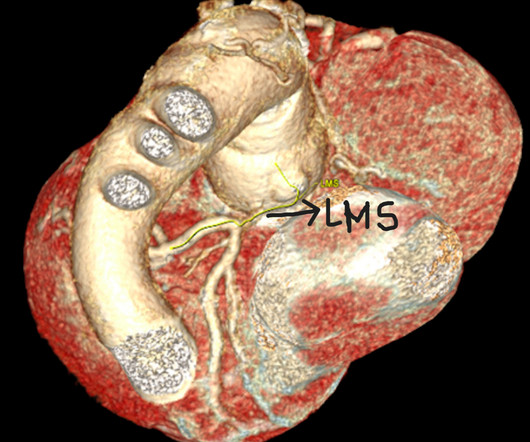
CT coronaryangiogram revealed severe narrowing of the left main coronary artery. Six months following modified Bentall procedure a patient presented with angina and acute ST depression. Minimal invasive off p.
His coronaryangiogram (CAG) is shown in figure 1 ( ). Figure 1 (A–C) Coronaryangiogram of left and right coronary arteries. (D) He had global left ventricular (LV) hypokinesia, severe LV dysfunction (LVEF 30%) and preserved thickness of LV and interventricular septum in echo. D) ECG taken in emergency room.
The coronaryangiogram was normal. The patient was managed medically and was referred to us in view of worsening symptoms with severe left ventricular dysfunction and moderate aortic regurgitation. Echocardiogram, CT aortogram and late gadolinium imaging of the aorta have been shown in figure 1.
We look directly at the coronary arteries using a cardiac CT scan. Subscribe now Cardiac CT There are two types of cardiac CT: CT Coronary Artery Calcium (CAC) Scan CT CoronaryAngiogram (CTCA). The CAC scan looks for deposits of calcium in the areas of the coronary arteries as a proxy marker for plaque.
Here it is: So we looked for the followup: Cath lab was activated per protocol and coronaryangiogram found no angiographic significant obstructive disease in the LAD, LCX, and RCA. Then he said: "No wonder the next EKG we recorded just before she left for the cath lab was normal."
Finally, do a coronaryangiogram Possible alternative to pacing is to give a beta-1 agonist to increase heart rate. Use Lidocaine instead (lidocaine prevents the PVCs which cause R on T, and does not prolong the QT.) Discontinue all QT proloning medications, including azithromycin 6. Dobutamine is an acceptable alternative.
1 In 320 patients, the availability of vFFR data led to a significant reduction in the number of vessels considered to have important disease and changed the management in 22% of cases when compared with their classification using angiography alone.
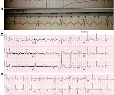
Will you accept this patient for emergent coronaryangiogram based on the ECG changes? The patient is a 70 something female with chest discomfort and dyspnea. How would you interpret the ST changes seen in this ECG? Does the ECG represent STEMI-negative OMI findings? How would you mange this patient?
Workup including routine laboratory results, 12-lead electrocardiogram (ECG), echocardiogram, and coronaryangiogram was non-specific. During the intravenous lacosamide infusion, the patient developed sudden cardiac arrest caused by ventricular arrhythmias necessitating resuscitation. 2893C>T, p.Arg965Cys) in the SCN5A gene.
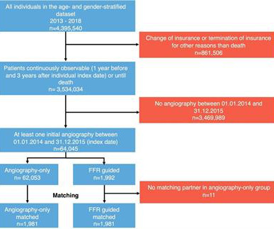
Patients undergoing coronary angiography between January 2014 and December 2015 were included in the analysis. Eligible patients had at least one inpatient coronaryangiogram for suspected coronary artery disease between January 2014 and December 2015.
The routine laboratory results, imaging study, coronaryangiogram, and echocardiogram (ECG) were normal. Case We report the case of a 20-year-old man with variants in SCN5A and RyR2 genes who was resuscitated from sudden cardiac death during sleep due to a ventricular fibrillation. The patient did not have underlying diseases.
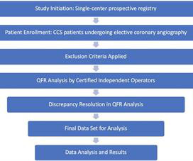
Methods This single-center prospective registry included consecutive patients with chronic coronary syndrome (CCS) who underwent elective coronary angiography, with or without revascularization.
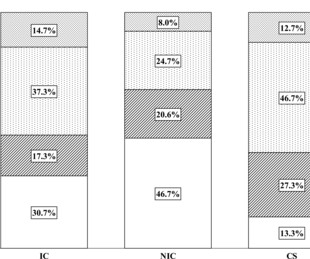
Objective The training of interventional cardiologists (ICs), non-interventional cardiologists (NICs) and cardiac surgeons (CSs) differs, and this may be reflected in their interpretation of invasive coronary angiography (ICA) and management plan.
Cardiology was consulted and the patient underwent coronaryangiogram which showed diffuse severe three-vessel disease. Coronaryangiogram shows diffuse severe three-vessel disease. Episodes of angina over past couple of months had been progressive. High sensitivity troponin I rose to peak at 2900 ng/L.
CT coronaryangiogram — No obstructive coronary disease. CT coronaryangiogram showed no obstructive coronary disease. But immediate resolution of chest pain once VT was converted — and — the normal CT coronaryangiogram — essentially ruled out acute coronary disease as the cause.
Her heart failure was due to the fistula as she had no coronary artery disease on coronaryangiogram. She had no further complications and was discharged home in good condition.Discussion:SVA affects the right sinus (94%), with the non-coronary (5%) and left sinus (1%) being less commonly involved.
Inclusion criteria included undergoing CAV screening with cardiac positron emission tomography scans and coronaryangiograms. Patients were grouped by the presence of angiographic CAV diagnosis and MBF reserve evaluated through cardiac positron emission tomography.
Written by Magnus Nossen The patient in todays case is a 50 year old male. He has a medical hx notable for hypertension, hyperlipidemia and previous tobacco use disorder. The patient presented due to chest pain that was typical in nature, retrosternal and radiating to the left arm and neck. He denied any exertional chest pain.
The cardiology fellow wrote in their note “unclear etiology of troponin elevation at this time, but hypertensive emergency, underlying CAD with demand ischemia , or NSTEMI all remain on the differential… In light of his risk factors, concerning clinical presentation and troponin trend -- we favor coronaryangiogram over CTCA at this time.”
Case:A 74-year-old male with a recent NSTEMI presented for elective coronary artery revascularization. We describe the "Double Guiding Catheter Technique" to minimize side branch closure and wire damage to non-atherectomy vessels during bifurcation orbital atherectomy (OA).Case:A
A repeat CT angiogram indicated contrast extension into the ventricular myocardium near the EPD but no lung spillage suggestive of pseudoaneurysm (Picture 1B).She A coronaryangiogram revealed normal arteries, while a left ventriculogram revealed contrast extravasation through the lateral wall (Picture 1C).
A Coronaryangiogram from 8 years prior revealed that he had had an inferior posterior STEMI at the time due to 100% occlusion of the proximal RCA. Regional wall motion abnormality- inferior and inferolateral.
addition to diagnostic coronaryangiogram, advances in noninvasive cardiac imaging allow further identification and characterization of these fistulae. Surgical approach was appropriate in this patient given the findings complex mvCAD and the CCF with significant hemodynamic shunt.
A repeat coronaryangiogram was unremarkable. Three hours later, a rise in CEs was noted, and EKG showed ST elevations inferiorly. TTE showed a reduced EF with multiple segmental abnormalities concerning for myocardial infarction. Intra-operative TEE showed an EF of 20% with no improvement after drainage.
Same catheter is used for engaging right and left coronary artery. Tiger catheter is the most popular catheter for transradial coronary angiography. Same catheter can be used for the right and left coronaryangiograms. Picture of a tiger diagnostic coronary catheter.
It’s judicious, then, to arrange for coronaryangiogram. Coronary occlusion, however, might be present concurrently with subendocardial ischemia on the time-zero ECG, or evolve into such. Proximal LAD disease with/without a) and b) It seemed quite apparent that this was an Acute Coronary Syndrome. CoronaryAngiogram 1.
All patients had CT coronaryangiograms at the start of the study and repeated after about one year. The DISCO trial is a more recent study that attempted to assess the role of nutrition in reversing plaque buildup 6. There was no increase in overall plaque in the intervention arm, but there was a small increase in the control arm.
Sleep & Heart Disease Cardiac CT - CAC Scores & CT CoronaryAngiograms. Understanding Blood Pressure. The Role of Inflammation. Formulas For Estimating Benefit & Risk. I'm Interested The Reducing Risk module is where we put it all together.
Images on the left panel show an example of coronary angiography, first-pass perfusion maps and stress and rest myocardial perfusion maps of a patient with cardiac amyloidosis. The coronaryangiogram is unobstructed and visual analysis of first-pass perfusion shows a global reduction in myocardial blood flow.
Outcome The only followup we got was that the patient is undergoing Coronary Bypass (CABG) of LAD, 2nd Obtuse Marginal, and Left Posterolateral coronaries. We don't know if he had a stress test, a CT Coronaryangiogram, or they just decided to do an angiogram.
Cardiology admitted him for observation with plans for next-day coronaryangiogram. Unfortunately, due to the patient’s abrupt exodus from the PCI center – without benefit of coronaryangiogram, or echo, for example – the disposition will forever remain unknown. [1] The peak Troponin I confirmed myocardial infarction. (A
We organize all of the trending information in your field so you don't have to. Join thousands of users and stay up to date on the latest articles your peers are reading.
You know about us, now we want to get to know you!
Let's personalize your content
Let's get even more personalized
We recognize your account from another site in our network, please click 'Send Email' below to continue with verifying your account and setting a password.


Let's personalize your content