This site uses cookies to improve your experience. To help us insure we adhere to various privacy regulations, please select your country/region of residence. If you do not select a country, we will assume you are from the United States. Select your Cookie Settings or view our Privacy Policy and Terms of Use.
Cookie Settings
Cookies and similar technologies are used on this website for proper function of the website, for tracking performance analytics and for marketing purposes. We and some of our third-party providers may use cookie data for various purposes. Please review the cookie settings below and choose your preference.
Used for the proper function of the website
Used for monitoring website traffic and interactions
Cookie Settings
Cookies and similar technologies are used on this website for proper function of the website, for tracking performance analytics and for marketing purposes. We and some of our third-party providers may use cookie data for various purposes. Please review the cookie settings below and choose your preference.
Strictly Necessary: Used for the proper function of the website
Performance/Analytics: Used for monitoring website traffic and interactions
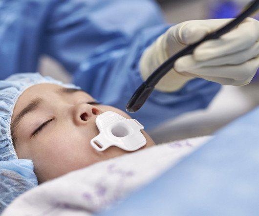
Cardiovascular ultrasound has played a key role in the evolution of early diagnosis of structural heart disease, led by a technology pioneered by Philips: the ‘transesophageal echocardiography’ (TEE) ultrasound transducer. TEE helps cardiologists by providing highly detailed images of the heart and its internal structures.
Image courtesy: Philips christine.book Wed, 06/12/2024 - 14:07 June 12, 2024 — Royal Philips has announced its next-generation AI-enabled cardiovascular ultrasound platform to help speed up cardiac ultrasound analysis with proven AI technology and reduce the burden on echocardiography labs.
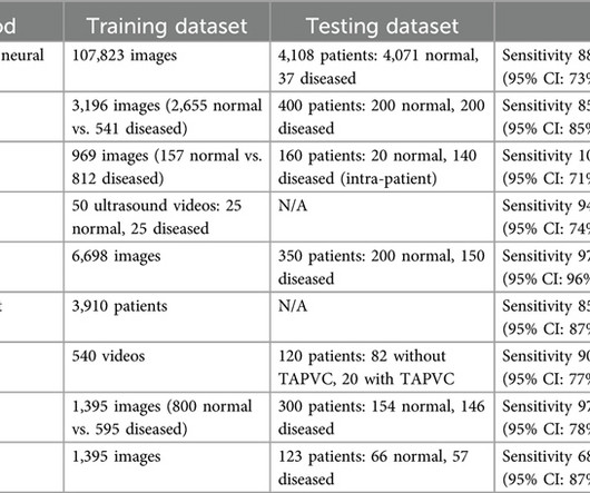
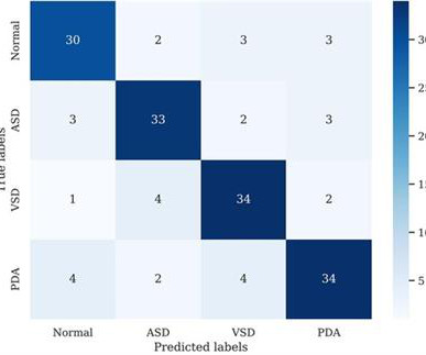
Artificial intelligence (AI)-powered ultrasound provides a potential solution to improve the diagnostic accuracy of fetal CHD screening.MethodsA literature search was conducted across seven databases for systematic review. Most studies utilized deep learning models using either ultrasound or echocardiographic images.
Food and Drug Adminstration (FDA) has approved DEFINITY (Perflutren Lipid Microsphere) as an ultrasound enhancing agent for use in pediatric patients with suboptimal echocardiograms, including those who have undergone heart transplant, or have Kawasaki disease or a congenital cardiovascular anomaly.
As far as congenital heart diseases go, HLHS falls on the rarer end of the spectrum. Zane was diagnosed during a routine ultrasound at 20 weeks’ gestation and his mom, Kayla, was immediately referred to the Colorado Fetal Care Center (CFCC) at Childrens Hospital Colorado.
Ultrasound techniques currently used in echocardiography uses frame rates from 30-150 frames/s. This limits its temporal resolution for very short lived events, especially in pediatric and congenital heart disease with faster heart rates compared to adults [1]. J Am Coll Cardiol. 2024 Jan 2;83(1):63-81. doi: 10.1016/j.jacc.2023.10.025.
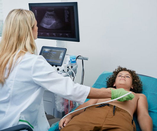
Congenital heart disease is a prevalent birth defect, accounting for approximately one-third of major birth defects. However, the emergence of deep learning in computer vision has paved the way for detecting subtle changes in chest x-rays, such as lung vessel density, enabling the detection of congenital heart disease in children.
Cardiac device therapy is frequently required for individuals with adult congenital heart disease (ACHD), whether it be for bradyarrhythmia, ventricular tachyarrhythmia, or cardiac resynchronization therapy (CRT).
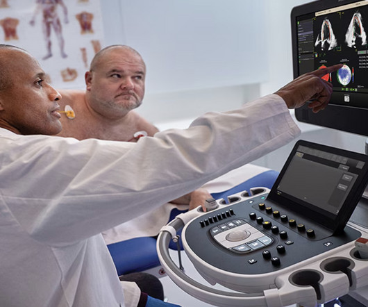
This year’s program, “Global Echocardiography: Innovations in Diagnosis and Beyond,” is described as an educational experience gathering global experts and enthusiasts in echocardiography, hosted by the largest global organization for cardiovascular ultrasound imaging serving physicians, sonographers, nurses, and scientists.
Purpose This study aims to evaluate deep learning (DL) denoising reconstructions for image quality improvement of Doppler ultrasound (DUS)-gated fetal cardiac MRI in congenital heart disease (CHD). Cine imaging was acquired using a balanced steady-state free precession (bSSFP) sequence with Doppler ultrasound gating.
Inferior vena cava (IVC) agenesis is a rare congenital anomaly that has been implicated in up to 5% of unprovoked deep vein thrombosis (DVT) cases in young men under 30 years old. A venous Doppler ultrasound revealed an extensive right lower extremity DVT.
Bedside cardiac ultrasound showed moderately decreased LV function. It should be kept in mind that on occasions, beta-one agonist can result in increased ventricular ectopy e.g., in severe myocardial ischemia (by increasing myocardial demand), or sometimes with congenital long-QT syndrome. She was intubated.
Functional single ventricle represents a complex congenital cardiac malformation where ventricular function directly impacts patients quality of life and prognosis. Accurate assessment of ventricular function.
Methods We retrospectively reviewed children with congenital heart diseases (CHDs) who received trans-axillary arterial catheterizations between January 2019 and February 2023. We aimed ultrasound-guided punctures in the proximal two-thirds of axillary arteries with diameters ≥2 mm to insert 7 cm/4 Fr short introducers.
We describe a fetus with prenatal echocardiographic findings of BDA and right aortic arch mirror-image branching (RAA-MIB) combined with congenital heart disease. Prenatal ultrasound diagnosis of BDA is important and requires a combination of 2D grayscale, CDFI, and STIC images to assist in scanning.
Introduction:Dextrocardia is a rare congenital condition where the heart's apex points to the right, with an incidence of about 0.01%. An intravascular ultrasound was also performed, which was negative for vessel dissection. Circulation, Volume 150, Issue Suppl_1 , Page A4140682-A4140682, November 12, 2024.
A bedside cardiac ultrasound was normal. Accelerated ventricular rhythm in children: a review and report of a case with congenital heart disease 3. There was apparently no syncope and he had no bony injuries, but he did complain of left sided chest pain. His chest was tender. He wrote: "ECG 1 - shows wide ???IVCD IVCD type rhythm ??
The heart is a pump and that pump may be faulty either due to congenital problem or an acquired problem. This is an ultrasound (a bit like the type that we use on pregnant women to look at the baby). Broadly speaking there are 3 things that can go wrong with the heart.
During echocardiography, a transducer transmits the ultrasound beam towards the heart. This becomes more difficult in complex congenital heart diseases where the cardiac chamber positions and size may vary. It is used in the emergency department, at bedside, in the intensive care unit as well as in the operating room.
A bedside cardiac ultrasound was performed with a parasternal long axis view demonstrated below: There is a large pericardial effusion with collapse of the right ventricle during systole. The beat-to-beat variation in QRS amplitude and morphology is electrical alternans. This patient is only pseudo-stable. She has already had syncope.
BackgroundThe Anomalous Origin of the Right Coronary Artery from the Left Coronary Sinus (ARCA-LCS) is a rare congenital cardiac condition where the right coronary artery emerges from the left sinus instead of the right coronary sinus of Valsalva. No previous history of hypertension or diabetes.
We organize all of the trending information in your field so you don't have to. Join thousands of users and stay up to date on the latest articles your peers are reading.
You know about us, now we want to get to know you!
Let's personalize your content
Let's get even more personalized
We recognize your account from another site in our network, please click 'Send Email' below to continue with verifying your account and setting a password.


Let's personalize your content