This site uses cookies to improve your experience. To help us insure we adhere to various privacy regulations, please select your country/region of residence. If you do not select a country, we will assume you are from the United States. Select your Cookie Settings or view our Privacy Policy and Terms of Use.
Cookie Settings
Cookies and similar technologies are used on this website for proper function of the website, for tracking performance analytics and for marketing purposes. We and some of our third-party providers may use cookie data for various purposes. Please review the cookie settings below and choose your preference.
Used for the proper function of the website
Used for monitoring website traffic and interactions
Cookie Settings
Cookies and similar technologies are used on this website for proper function of the website, for tracking performance analytics and for marketing purposes. We and some of our third-party providers may use cookie data for various purposes. Please review the cookie settings below and choose your preference.
Strictly Necessary: Used for the proper function of the website
Performance/Analytics: Used for monitoring website traffic and interactions
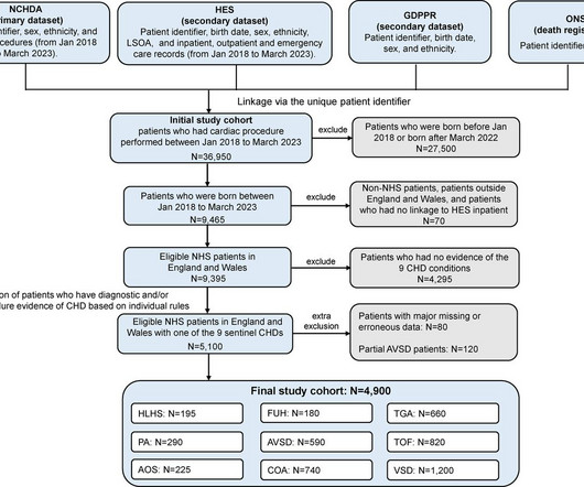
Background Infants with congenital heart disease (CHD) are clinically vulnerable to cardiac deteriorations and intercurrent infections. We aimed to quantify the impact of health system disruptions during the COVID-19 pandemic, on their clinical outcomes and whether these differed by socioeconomic and ethnic subgroups.
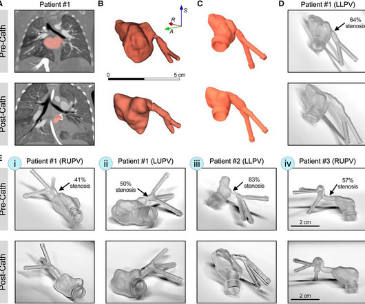
IntroductionPrimary pulmonary vein stenosis (PVS) is a rare congenital heart disease that proves to be a clinical challenge due to the rapidly progressive disease course and high rates of treatment complications. These 3D reconstructions were 3D printed using a clear resin ink and used in a benchtop experimental setup.
Tetralogy of Fallot TOF with pulmonary atresia Pulmonary atresia with intact interventricular septum Tricuspid atresia Double outlet right ventricle Transposition of great arteries with ventricular septal defect and pulmonarystenosis Ebstein’s anomaly of tricuspid valve In DORV and tricuspid atresia, there are also variants with increased pulmonary (..)
D-Transposition of great arteries Double outlet right ventricle without pulmonarypulmonarystenosis Taussig-Bing anomaly Total anomalous pulmonary venous return Truncus arteriosus Single ventricle (double inlet ventricle, univentricular heart)
Transcatheter pulmonary valve replacement (TPVR) has become a safe and effective alternative to surgical PVR in tetralogy of Fallot (TOF), isolated pulmonarystenosis (PS), and other congenital heart disease (CHD) variants.
Tetralogy of Fallot (TOF) is the most common form of cyanotic congenital heart disease (CHD). Early management of this condition is typically dictated by the degree of pulmonarystenosis (PS) and resulting oxygen saturations.
The desired ultimate ability for such devices to treat a vascular stenosis without long-term device-related complications or impeding future treatment continues to evoke excitement in clinicians and engineers alike.
Animal studies have shown that mice with TBX1 gene mutations have smaller left pulmonary arteries compared to wild type mice, defined by a reduced left pulmonary artery (LPA) to right pulmonary artery (RPA) ratio. A single study has shown this translates to humans with 22q11 and structurally normal hearts.
Right Heart Catheterization in Tetralogy of Fallot With the availability of high resolution echocardiographic images and Doppler echocardiography, role of cardiac catheterization has come down in tetralogy of Fallot and other congenital heart diseases in general. Normal subjects have a value around 2.1. If McGoon’s ratio is below 0.8,
Stenotic lesions included 16 branch pulmonary arteries, 9 aortic isthmus, 2 right ventricular outflow tracts, and 1 Glenn anastomosis. Percentage of stenosis was 50% (IQR, 36%58%). mm (IQR, 7.59.5) (P<0.001), with median stenosis expansion at 103% (IQR, 51%146%). male) with median age and weight of 3.4 kg (IQR, 9.116.4).
Due to the dramatically improved survival of children with congenital heart disease over the last 5 decades, there has been a steady increase in the prevalence of adults with congenital heart disease, which necessitates that clinicians become familiar with the anatomy and the evaluation of right ventricular outflow tract and PV anomalies.
Abstract Introduction Patients with congenital heart disease are at increased risk for requiring cardiac pacing during their lifetime. Due to atrial and ventricular pacing dependence, a comprehensive congenital care team concluded the need for lead extraction and replacement of pacemaker via leadless peacemaking device.
We know a small ASD decompresses mitral stenosis, and the combination of ASD and MS, Lutembacher, is a well-known syndrome called Lutembacher. This is to create a small regulatory orifice in the IAS ( A complicated term for a small ASD ) to decompress the LA and reduce pulmonary congestive symptoms. JAMA Cardiol.
The Y descent is shallow in tricuspid stenosis, and absent in cardiac tamponade. Right atrial hypertrophy as in tricuspid stenosis, pulmonarystenosis and pulmonary hypertension. But in a VSD with pulmonary hypertension A wave is not prominent. X descent, X prime descent and Y descent.
This becomes more difficult in complex congenital heart diseases where the cardiac chamber positions and size may vary. The aorta, right ventricular outflow tract and pulmonary artery up to its bifurcation is imaged in the upward angulation shown in the left panel. Colour flow shows the flow in pulmonary artery.
BackgroundPulmonary vein stenosis in children is associated with a poor prognosis. However, the cause and risk factors for mortality remain uncertain.MethodsThis retrospective, singlecenter study identified children with primary and secondary pulmonary vein stenosis through a cardiac catheterization database. 95% CI, 2.214.1];P<0.001).
Transcript of video: Tetralogy of Fallot is one of the commonest cyanotic congenital heart diseases. One is ventricular septal defect, second is overriding aorta, third is pulmonarystenosis, usually right ventricular outflow tract stenosis and associated right ventricular hypertrophy. Right to left shunt is also visible.
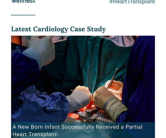
In a remarkable leap forward for pediatric cardiac care, a groundbreaking partial heart transplant procedure has emerged as a beacon of hope for infants facing severe congenital heart conditions. The surgical team skillfully dissected the pulmonary artery ostia and coronary artery buttons, preparing for the intricate transplant.
On the other hand, the murmur in valvular aortic stenosis does not change substantially or decreases slightly following the Valsalva maneuver. A decrease in intensity, due to attenuation of obstruction, is heard after going from a standing to a sitting or squatting position, with a handgrip, and following passive elevation of the legs.
severe mitral stenosis, pulmonary hypertension, or cardiomyopathy), prolonged labor could strain the heart excessively, potentially leading to decompensation, heart failure, or arrhythmias. repaired congenital defects) might safely attempt vaginal delivery with careful monitoring and a low threshold for intervention.
We organize all of the trending information in your field so you don't have to. Join thousands of users and stay up to date on the latest articles your peers are reading.
You know about us, now we want to get to know you!
Let's personalize your content
Let's get even more personalized
We recognize your account from another site in our network, please click 'Send Email' below to continue with verifying your account and setting a password.


Let's personalize your content