This site uses cookies to improve your experience. To help us insure we adhere to various privacy regulations, please select your country/region of residence. If you do not select a country, we will assume you are from the United States. Select your Cookie Settings or view our Privacy Policy and Terms of Use.
Cookie Settings
Cookies and similar technologies are used on this website for proper function of the website, for tracking performance analytics and for marketing purposes. We and some of our third-party providers may use cookie data for various purposes. Please review the cookie settings below and choose your preference.
Used for the proper function of the website
Used for monitoring website traffic and interactions
Cookie Settings
Cookies and similar technologies are used on this website for proper function of the website, for tracking performance analytics and for marketing purposes. We and some of our third-party providers may use cookie data for various purposes. Please review the cookie settings below and choose your preference.
Strictly Necessary: Used for the proper function of the website
Performance/Analytics: Used for monitoring website traffic and interactions
Echocardiograms using the robotic arm resulted in the same diagnosis as conventional in-person echocardiography in 98% of cases (papillary muscle level obstruction was missed in one case). tim.hodson Thu, 08/29/2024 - 11:39 Aug. 28, 2024 — New research presented at this year’s ESC Congress 2024 in London, UK (Aug. 30 – Sept.
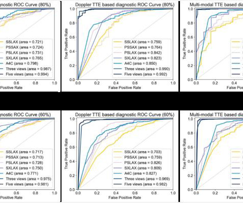
milla1cf Wed, 05/29/2024 - 07:00 May 29, 2024 — Congenital heart disease (CHD) is one of the most common congenital anomalies worldwide, which brings a heavy health and financial burden to patients. However, inexperienced sonographers often face difficulties in recognizing CHD through transthoracic echocardiogram (TTE) images.
Food and Drug Adminstration (FDA) has approved DEFINITY (Perflutren Lipid Microsphere) as an ultrasound enhancing agent for use in pediatric patients with suboptimal echocardiograms, including those who have undergone heart transplant, or have Kawasaki disease or a congenital cardiovascular anomaly. and Description (11)].
Objective To evaluate for correlation between exercise capacity as assessed by peak oxygen consumption (pVO 2 ) measurement during a cardiopulmonary exercise test (CPET) and smartwatches reporting this parameter in patients with adult congenital heart disease (ACHD) complex lesions. Apple Watch was the predominant smartwatch used (79%).
It should be kept in mind that on occasions, beta-one agonist can result in increased ventricular ectopy e.g., in severe myocardial ischemia (by increasing myocardial demand), or sometimes with congenital long-QT syndrome. Therefore, I usually prefer temporary pacing which might be more controlled and is more predictable." J Am Coll Cardiol.
BackgroundThe complexity of congenital heart disease has been primarily stratified on the basis of surgical technical difficulty, specific diagnoses, and associated outcomes. The panel refined complexity categories and included study modifiers to account for complexity related to performance of the echocardiogram. were outpatient; 34.5%
In preparation for the ABIM Cardiovascular Disease exam, check out the BoardVitals Cardiology Board Review Question Bank and we’ll make sure you’re well versed in the following 13 areas covered on the exam: Multiple-Choice Component Arrhythmias 15% Coronary Artery Disease 23% Heart Failure and Cardiomyopathy 17% Valvular Disease 15% Pericardial (..)
Abstract Introduction Patients with congenital heart disease are at increased risk for requiring cardiac pacing during their lifetime. Due to atrial and ventricular pacing dependence, a comprehensive congenital care team concluded the need for lead extraction and replacement of pacemaker via leadless peacemaking device.
of all congenital cardiac anomalies. Her ejection fraction was 66% ejection fraction with a fistula between the right sinus of Valsalva and the right atrium on transthoracic echocardiogram (TTE) which was also seen on transesophageal echocardiogram (TEE). Introduction:Sinus of Valsalva aneurysm (SVA) accounts for 3.5%
On his bib it stated that he had a congenital heart disorder. An echocardiogram confirmed aortic stenosis with a large pressure gradient. This young male had ventricular fibrillation during a triathlon. He was resuscitated with chest compressions and defibrillation and 1 mg of epinephrine. His initial ECG is shown here.
The LPA to RPA ratio on initial and most recent echocardiogram, intervention on the LPA at initial surgery and subsequent reintervention on the LPA were compared between the two groups.Results:The 22q11 deletion and control group had a similar mean age at time of study 6.9±3.4 0.27, p=0.002) echocardiogram.
Right Heart Catheterization in Tetralogy of Fallot With the availability of high resolution echocardiographic images and Doppler echocardiography, role of cardiac catheterization has come down in tetralogy of Fallot and other congenital heart diseases in general. Magnetic resonance imaging is another way of documenting coronary anomalies.
After excluding patients with congenital or rheumatic heart disease, heart transplant recipients, or those without baseline echocardiogram, a total of 130 patients were included in the analysis. Echocardiographic data were analyzed at baseline before ablation, and at early follow-up within 1-year postablation.
Introduction:Dextrocardia is a rare congenital condition where the heart's apex points to the right, with an incidence of about 0.01%. Circulation, Volume 150, Issue Suppl_1 , Page A4140682-A4140682, November 12, 2024. Patients usually have a normal life expectancy unless other structural heart diseases are present.
Similarly, for echocardiogram, what we would do usually is, first we do a clinical history evaluation, then physical examination, and after that only we proceed with echocardiography in our routine work. You can see the two dimensional sector imaging from an echocardiogram and I have marked out the aorta.
The image shown here is an animated 2 dimensional echocardiogram. This one is an older mode known as time-motion mode or M-Mode echocardiogram. This becomes more difficult in complex congenital heart diseases where the cardiac chamber positions and size may vary. First view to be obtained is often the parasternal long axis view.
The heart is a pump and that pump may be faulty either due to congenital problem or an acquired problem. Overall though a normal cardiac MRI is even more reassuring than a normal echocardiogram. Broadly speaking there are 3 things that can go wrong with the heart. A simple standard 12 lead ECG can be helpful.
Congenital Heart Defects Some individuals are born with heart defects that cause parts of the heart to work harder, leading to enlargement over time. Echocardiogram An echocardiogram uses sound waves to produce a detailed image of the heart, allowing doctors to see the size of the heart chambers and how well the heart is pumping blood.
Model performance was evaluated on single ECG–echocardiogram pairs per patient at Boston Children’s Hospital and externally at Mount Sinai Hospital using area under the receiver operating characteristic curve (AUROC) and area under the precision-recall curve (AUPRC).RESULTS:The composite outcomes) cohorts.
However, an echocardiogram is a different test, also conducted for heart activity. Hypoplastic left heart syndrome is a common congenital heart defect in which there is a problem with the heart's blood supply, and the left side of the heart does not develop correctly. ECG and EKG refer to the same thing.
Echocardiogram is indicated (Correct) C. Start aspirin and Plavix Correct answer: (B) (B) Echocardiogram is indicated. Which of the following is the best statement to describe further clinical management? No further workup is indicated B. Start furosemide for diuresis D.
An echocardiogram revealed the heart condition that would define much of his early life: severe congenital heart defects. At birth, he was small, which led to a car seat test being ordered to ensure his safety. That test revealed an irregular heart rate, prompting a more in-depth evaluation.
BackgroundThe Anomalous Origin of the Right Coronary Artery from the Left Coronary Sinus (ARCA-LCS) is a rare congenital cardiac condition where the right coronary artery emerges from the left sinus instead of the right coronary sinus of Valsalva. No previous history of hypertension or diabetes.
We organize all of the trending information in your field so you don't have to. Join thousands of users and stay up to date on the latest articles your peers are reading.
You know about us, now we want to get to know you!
Let's personalize your content
Let's get even more personalized
We recognize your account from another site in our network, please click 'Send Email' below to continue with verifying your account and setting a password.


Let's personalize your content