This site uses cookies to improve your experience. To help us insure we adhere to various privacy regulations, please select your country/region of residence. If you do not select a country, we will assume you are from the United States. Select your Cookie Settings or view our Privacy Policy and Terms of Use.
Cookie Settings
Cookies and similar technologies are used on this website for proper function of the website, for tracking performance analytics and for marketing purposes. We and some of our third-party providers may use cookie data for various purposes. Please review the cookie settings below and choose your preference.
Used for the proper function of the website
Used for monitoring website traffic and interactions
Cookie Settings
Cookies and similar technologies are used on this website for proper function of the website, for tracking performance analytics and for marketing purposes. We and some of our third-party providers may use cookie data for various purposes. Please review the cookie settings below and choose your preference.
Strictly Necessary: Used for the proper function of the website
Performance/Analytics: Used for monitoring website traffic and interactions
Circulation: Cardiovascular Imaging, Ahead of Print. BACKGROUND:Recently, it was reported that noncalcified plaque (NCP) volume was an independent predictor for cardiac events. P<0.001) than the group with low NCP plaque volume. P<0.001) than the group with low NCP plaque volume. versus 75.9%;P<0.001),
The early detection of plaques by circulating biomarkers is highly clinically relevant to prevent the occurrence of major complications such as stroke or heart attacks. miRNA expression profiles of serum-derived EVs were obtained by small RNA sequencing and in plaque material simultaneously acquired from patients.
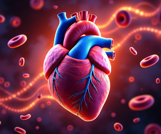
This is where coronary circulation comes into play. Coronary circulation refers to the movement of blood through the network of coronary arteries and veins that supply the heart muscle (myocardium) itself. Step-by-Step Breakdown of Coronary Circulation 1. Any interruption in this process can result in serious consequences.
Thus, it has recently become generally accepted that most plaque ruptures resulting in myocardial infarction occur in plaques that narrow the lumen diameter by 40% of the arterial cross section may be involved by plaque. The pathologist may see a plaque that constitutes, for example, 50% of the cross-sectional area.
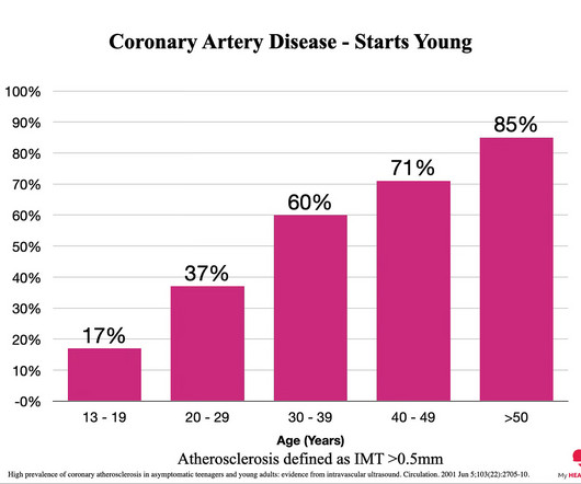
Everyone starts with no plaque in the coronary arteries, but over a long enough time frame, everyone develops plaque in their coronary arteries. By age 80, almost everyone will have evidence of advanced plaque in their coronary arteries, as defined by a cardiac CT 1. Plaque accumulation happens in stages. You got it.
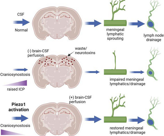
Skull development coincides with the onset of cerebrospinal fluid (CSF) circulation, brain-CSF perfusion, and meningeal lymphangiogenesis, processes essential for brain waste clearance. Further, craniosynostosis exacerbates amyloid pathology and plaque buildup in Twist1+/–:5xFAD transgenic Alzheimer’s disease models.
Introduction:Nonstenotic carotid plaque is found in some patients with otherwise cryptogenic stroke (CS) but also in normal individuals and patients with stroke of known cause (KS). In formal meta-analysis, the summary risk ratio for nonstenotic carotid plaque in CS vs KS patients was 1.54 (95%CI 1.15-2.05) 2.05) (Figure).
Circulation, Volume 150, Issue Suppl_1 , Page A4144040-A4144040, November 12, 2024. The sections were also used for immunohistochemistry with cell marker antibodies (to measure % plaque smooth muscle cells (SMC), macrophages (MF), and endothelial cells (EC)). to D: 30.7%), elevation in plaque MF (A: 16.7%
The reason: They were accumulating plaque in their coronary arteries much earlier than their peers. You can’t have a heart attack if you don’t have plaque in your coronary arteries. And plaque in your coronary arteries is the result of exposure to risk factors over time. The answer: Risk Factors. The answer.
If the arrest was caused by acute MI due to plaque rupture, then the diagnosis is MINOCA. Here is my comment on MINOCA: "Non-obstructive coronary disease" does not necessarily imply "no plaque rupture with thrombus." They often cannot even be recognized as culprits, as fissured or ulcerated plaque. FFR can be useful.
Introduction:A new Plaque-RADS classification (I-IV) is proposed to categorize the degree of carotid plaque instability and risk of embolic ischemic stroke. Carotid total plaque thickness and ulceration were scored by a neuroradiologist blinded to stroke side. N=188 plaques) met criteria.
Circulation: Cardiovascular Imaging, Ahead of Print. A decrease in the percentage of unstable core (fibro-fatty+necrotic plaque; from 14.1 [7.9–22.3] BACKGROUND:Intensive lipid-lowering therapy may induce coronary atherosclerosis regression. The global coronary PB changed from 34.6% (32.5%–36.8%) 36.8%) at entry to 30.4% (27.4%–33.4%)
MINOCA may be due to: coronary spasm, coronary microvascular dysfunction, plaque disruption, spontaneous coronary thrombosis/emboli , and coronary dissection; myocardial disorders, including myocarditis, takotsubo cardiomyopathy, and other cardiomyopathies. Circulation [Internet] 2017;135(16):1481–9. Circulation. From Gue at al.
Non-obstructive coronary disease at the time cardiac cath is done does not necessarily imply there was no plaque rupture with thrombus. These plaques will often not be recognized as "culprits" — because no fissuring or ulceration is seen.
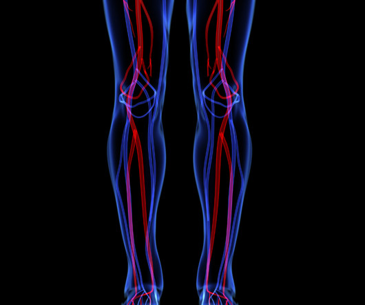
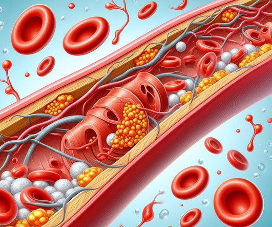
PAD is a serious condition affecting circulation and blood vessels, causing them to narrow from plaque buildup in the arteries and blocking blood flow to the extremities, typically the legs and feet. It is estimated that more than 10 million Americans are living with PAD, or Peripheral Artery Disease.
PAD occurs when plaque builds up in the arteries and limits blood flow to the lower extremities. This limited blood flow causes poor circulation, which if left un-treated, can severely damage foot and leg tissue, sometimes resulting in non-healing ulcers.
Specific genetic variants, such as those affecting cholesterol metabolism, can increase the likelihood of plaque buildup in the arteries. Exercise: Regular physical activity strengthens the heart and improves blood circulation. Cardiomyopathies: These diseases affect the heart muscle, impairing its ability to pump blood effectively.
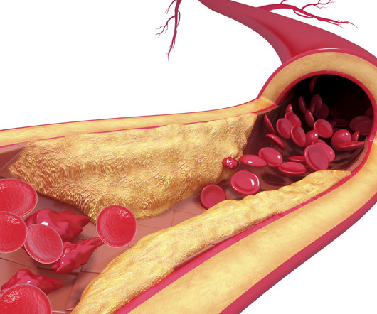
When we say heart disease, what we really mean is plaque in the artery wall. Therefore, the number of ApoB particles is a direct measure of the number of circulating cholesterol particles in your bloodstream. You just need to know how. This Is Known As Atherosclerosis. We also refer to these cholesterol particles as ApoB particles.
Lp(a) is emerging as an important, yet under-recognized, potential risk factor for cardiovascular disease due to its ability to promote the development of plaques within artery walls, clot formation and aortic valve calcification. Circulation. Arnett DK et al. 2019 Sep 10;140(11):e563-e595. doi: 10.1161/CIR.0000000000000677.
However, most adults will start to develop advanced plaque in their coronary arteries early in life. By age 66, more than half of all females will have evidence of advanced plaque in their coronary arteries, as seen on a CT calcium score. Coronary atherosclerosis, as evidenced by an abnormal CAC score, is a measure of advanced plaque.
people from the general population), coronary artery calcium scores (CACS) are higher, indicating more calcification and the presence of atherosclerotic plaques. Calcified plaques are known to be more stable and less prone to rupture and lead to a heart attack. When comparing athletes to control groups (i.e., hours per week).
Circulation, Ahead of Print. Sections of coronary atherosclerotic plaques from donors were immunostained to analyze calcium deposition and 4-HNE. Vascular calcification is associated with increased cardiovascular adverse events.
Atherosclerotic cardiovascular disease (ASCVD), caused by plaque buildup in arterial walls, is one of the leading causes of disability and death worldwide.1,2 7 Research has shown inflammation plays a significant role in the development of atherosclerosis and ASCVD,8-10 and even the formation of plaque.11 Circulation.
Circulation, Volume 150, Issue Suppl_1 , Page A4142898-A4142898, November 12, 2024. The primary outcome was the prevalence of carotid artery plaques assessed by ultrasound. Secondary outcomes included changes in NMR-derived lipoprotein subclasses and their mediation effects on carotid plaque. day) or low-dose (1.5g/day)
Atherosclerotic plaque formation is modulated by genetic and environmental interactions. In this study, we examined the effects of ApoE deficiency and high fat diet (HFD) on plaque formation and immunomodulatory cellular and molecular mechanisms in mice models.Methods:Wild type and ApoE knockout (KO) mice were fed with normal or HFD.
Circulation, Volume 150, Issue Suppl_1 , Page A4140066-A4140066, November 12, 2024. The incidence of no-reflow was higher in patients with attenuated plaque ≥5 mm in length as evaluated by intravascular ultrasound (IVUS).Objective:The Blood pressure decrease during PCI was significantly more pronounced in the no-reflow group (47.4%
Circulation, Ahead of Print. BACKGROUND:Mitochondrial dysfunction is a key factor in the development of atherogenesis. METTL4 (methyltransferase-like protein 4) mediates N6- methyldeoxyadenosine (6mA) of mammalian mitochondrial DNA (mtDNA). However, the role of METTL4-mediated mitoepigenetic regulation in atherosclerosis is still unknown.
Circulation, Volume 150, Issue Suppl_1 , Page A4132968-A4132968, November 12, 2024. M-MDSCs phenotype switch, atherosclerotic lesion development and plaque phenotype was studiedin vivo.Results:We observed CCR5 elevation on Monocytic-MDSCs in the early phase of atherosclerosis. 3ng/day for 15 days).M-MDSCs
Circulation: Genomic and Precision Medicine, Ahead of Print. 10−4, we identified 3 genes (ATP1B1,ARVCF, andLIPG) associated with CAC and 2 genes (ABCG8andEIF2B2) associated with carotid intima-media thickness and carotid plaque, respectively, through gene-based rare variant set analysis.
Circulation, vol. It provides anatomic data, plaque identification and characterization, as well as the calculations of FFR CT , a coronary physiological simulation, computed from simulated pressure, velocity and blood flow information obtained from a 3D computer model generated from static coronary CT images. The Lancet, vol.
Circulating immune cells have been found to induce immunothrombosis, and they actively participate in the formation of the thrombus by promoting platelet recruitment and thrombin activation. Additionally, the formation of thromboinflammation leads to increased instability of atherosclerotic plaques.
Circulation, Volume 151, Issue 6 , Page 400-415, February 11, 2025. Lp(a) can enter the vessel wall, leading to the accumulation of oxidized phospholipids in the arterial intima, which are crucial for initiating plaque inflammation and triggering vascular disease progression.
However ,we have some effective clinical and pathological markers too, for effective re-vascularisation They are clinical well being and good functional capacity , relief from chest-pain, reduction of plaque volume, plaque stabilisation, maintenance of collaterals , microvascular patency , reduction of recurrent events.
Circulation, Volume 149, Issue 3 , Page 251-266, January 16, 2024. Nevertheless, the relationship between CAC and the susceptibility of a plaque to provoke a thrombotic event remains incompletely understood. Coronary artery calcification (CAC) accompanies the development of advanced atherosclerosis.
Coronary Artery Disease (CAD) CAD, which involves the narrowing or blockage of coronary arteries due to plaque buildup, can reduce blood flow to the heart. Regular physical activity can strengthen the heart and improve circulation. Avoid smoking and limit alcohol intake. Monitor blood pressure and cholesterol levels regularly.
They shocked him twice before return of spontaneous circulation. His ECG at the accepting facility is shown below: Accepting facility ECG The team reviewed his angiography films with an interventionalist and thought they were suspicious for plaque rupture in LAD, but they were not confident.
Circulation, Volume 148, Issue 24 , Page 1958-1973, December 12, 2023. Parabiosis studies revealed that monocyte-derived macrophages were significantly increased in the hearts of PCOS mice because of enhanced circulating Ly6C+monocyte supply. BACKGROUND:Reducing cardiovascular disease burden among women remains challenging.
To prove there is no plaque rupture, you need to do intravascular ultrasound (IVUS). An angiogram is a "lumenogram;" most plaque is EXTRALUMINAL!! One of the most common is rupture of a non-obstructive plaque, with thrombus formation and OMI that spontaneously lyses and leaves a wide open artery. It can only be seen by IVUS.
Circulation: Cardiovascular Interventions , 7(5), 645–655. Learning Points: Type 1 MI is the type we are most familiar with: rupture of atherosclerotic plaque with production thrombus or platelet fibrin aggregates. Buller, C. Starovoytov, A., Robinson, S., Vuurmans, T., Humphries, K., & & Mancini, G.
Circulation, Volume 150, Issue Suppl_1 , Page A4142716-A4142716, November 12, 2024. Background:Heavily calcified coronary bifurcation lesions present significant challenges during percutaneous coronary intervention, particularly during atherectomy due to the risk of side branch occlusion from plaque shift.
We organize all of the trending information in your field so you don't have to. Join thousands of users and stay up to date on the latest articles your peers are reading.
You know about us, now we want to get to know you!
Let's personalize your content
Let's get even more personalized
We recognize your account from another site in our network, please click 'Send Email' below to continue with verifying your account and setting a password.


Let's personalize your content