This site uses cookies to improve your experience. To help us insure we adhere to various privacy regulations, please select your country/region of residence. If you do not select a country, we will assume you are from the United States. Select your Cookie Settings or view our Privacy Policy and Terms of Use.
Cookie Settings
Cookies and similar technologies are used on this website for proper function of the website, for tracking performance analytics and for marketing purposes. We and some of our third-party providers may use cookie data for various purposes. Please review the cookie settings below and choose your preference.
Used for the proper function of the website
Used for monitoring website traffic and interactions
Cookie Settings
Cookies and similar technologies are used on this website for proper function of the website, for tracking performance analytics and for marketing purposes. We and some of our third-party providers may use cookie data for various purposes. Please review the cookie settings below and choose your preference.
Strictly Necessary: Used for the proper function of the website
Performance/Analytics: Used for monitoring website traffic and interactions
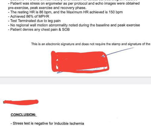
Post cath ECG: Now there are hyperacute T-waves again, and recurrent ST depression in V2 This ECG would normally diagnostic of OMI until proven otherwise No further troponins were measured, but it looks like there is recurrent OMI Next day: A CT CoronaryAngiogram was done (CTCA) CARDIAC MORPHOLOGY AND FUNCTION: 1. Circulation.
The diagnostic coronaryangiogram identified only minimal coronary artery disease, but there was a severely calcified, ‘immobile’ aortic valve. Aortic angiogram did not reveal aortic dissection. The ECG cannot diagnose the etiology of ischemia; it only the presence of ischemia, from whatever etiology.
We know, stress tests can give false positive results suggesting ischemia in at least 20% of patients for various reasons. Mostly, you can’t escape from a coronaryangiogram” Next option is CT angiogram, Thallium or dobutamine stress. The patient seeked by advice “It was indeed an academic stress test.
The patient was started on heparin for possible NSTEMI vs demand ischemia. increasing stenosis, ischemia, volume changes, increased blood pressure, atrial fibrillation, etc.) The EKGs from the ED presentation were felt by cardiology to represent "subendocardial ischemia." Smith : these ECGs do NOT show subendocardial ischemia.
That said there were no clinical symptoms or ECG findings suggestive of ongoing ischemia. CT coronaryangiogram showed a hypoplastic RCA and dominant LCx. You have given IV MgSO4 a fast acting -blocker and IV amiodarone bolus and infusion. Troponin T was negative on admission and on repeat blood draw. No PVCs are seen.
We organize all of the trending information in your field so you don't have to. Join thousands of users and stay up to date on the latest articles your peers are reading.
You know about us, now we want to get to know you!
Let's personalize your content
Let's get even more personalized
We recognize your account from another site in our network, please click 'Send Email' below to continue with verifying your account and setting a password.


Let's personalize your content