This site uses cookies to improve your experience. To help us insure we adhere to various privacy regulations, please select your country/region of residence. If you do not select a country, we will assume you are from the United States. Select your Cookie Settings or view our Privacy Policy and Terms of Use.
Cookie Settings
Cookies and similar technologies are used on this website for proper function of the website, for tracking performance analytics and for marketing purposes. We and some of our third-party providers may use cookie data for various purposes. Please review the cookie settings below and choose your preference.
Used for the proper function of the website
Used for monitoring website traffic and interactions
Cookie Settings
Cookies and similar technologies are used on this website for proper function of the website, for tracking performance analytics and for marketing purposes. We and some of our third-party providers may use cookie data for various purposes. Please review the cookie settings below and choose your preference.
Strictly Necessary: Used for the proper function of the website
Performance/Analytics: Used for monitoring website traffic and interactions
We used carotid ultrasounds to detect plaque at baseline and follow‐up in 2006 to 2009 (median follow‐up=5.5 had low‐density lipoprotein cholesterol ≥160 mg/dL, which is higher than the recommended threshold for lifestyle or medical interventions in young adults of 20 to 39 years old. Lipids were measured after a 12‐hour fast.
Plaque regression can be demonstrated by ultrasound evaluation of the carotids which are easily accessible. High density lipoprotein cholesterol in the blood increases with regular exercise. HDL is involved in reverse cholesterol transport from the blood vessels to the liver.
The primary outcome was the prevalence of carotid artery plaques assessed by ultrasound. Genetic interactions with n-3 PUFA supplementation were also analyzed.Results:There were 383 participants (92.3%) completed the 14-month intervention and 358 patients (86.3%) completed carotid ultrasound exams.
The fundamental characteristic of atherosclerosis is when a cholesterol particle becomes trapped in the artery wall. Each of these particles contains varying amounts of LDL cholesterol (CE) and triglycerides INSIDE the particle. CE = Cholesterol Ester. Share But what about LDL cholesterol? TG = Triglycerides.
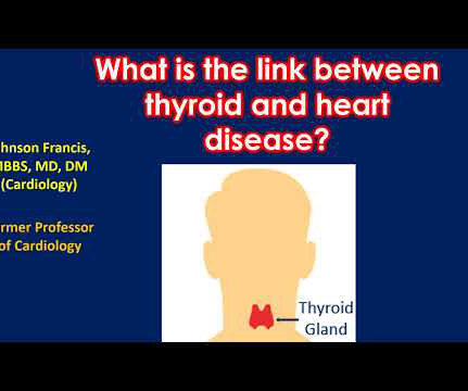
Cholesterol levels go up when thyroid function comes down. Ultrasound image of the heart – echocardiogram, showing fluid collection around the heart, marked as PE, short for pericardial effusion. Hence the blood stagnates in some parts of the upper chambers (left atrium) and clots may form.
Carotid ultrasound results were divided into two groups based on the presence or absence of plaque. This association remains independent even after adjustment for factors such as statin use or low-density lipoprotein cholesterol (LDL-C) levels. Lp(a) levels were categorized into two groups: below 50 mg/dl and 50 mg/dl or higher.
BACKGROUND:Small dense low-density lipoprotein cholesterol (sdLDL-C) particles are more atherogenic than large and intermediate low-density lipoprotein cholesterol (LDL-C) subfractions. Incident carotid plaques and their vulnerability were detected by carotid ultrasound at follow-up (2021). Stroke, Ahead of Print.
You cannot eliminate the plaque entirely, but multiple clinical trials have shown plaque regression using high-intensity cholesterol-lowering treatments, which I have discussed previously. 4 Coronary atheroma regression and plaque characteristics assessed by grayscale and radiofrequency intravascular ultrasound after aerobic exercise.
Few of them currently have the equipment and expertise to diagnose valvular heart disease, but recent studies have demonstrated that healthcare professionals can use a combination of portable ultrasound devices and AI to diagnose heart diseases as well.
Controlling LDL cholesterol. Let's take a 42-year-old male with an LDL cholesterol of 5.1 How much would they benefit from going on a cholesterol lowering medication? Now add in an elevated Lp(a), which is a common genetic cholesterol particle disorder, and that risk reduction gets even bigger. Being active. Not smoking.
It is also very important to mention a history of high blood pressure, diabetes, elevated cholesterol, family history of premature heart disease, stroke or even sudden death. Ultrasound – this is easily available, very portable and usually a very low risk investigation. There are a variety of ways to look at these.
Echocardiography – We can use ultrasound to visualize the heart and look at how well it pumps. With time, fat and cholesterol can get trapped in the areas of wear and tear and cause plaque formation. This is termed as diastolic dysfunction. So what tests tell us about the heart as a pump? The plaques can damage us in 2 ways.
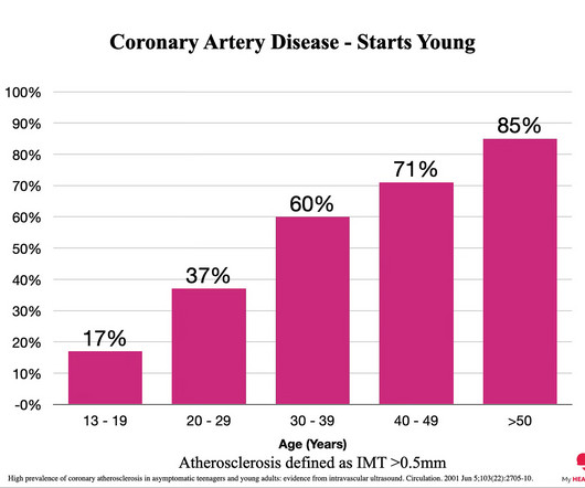
These noninvasive scans look directly at the coronary arteries rather than assessing for the risk factors for coronary artery disease eg LDL cholesterol, high blood pressure etc. 4 High prevalence of coronary atherosclerosis in asymptomatic teenagers and young adults: evidence from intravascular ultrasound. CT Coronary Angiogram.
We organize all of the trending information in your field so you don't have to. Join thousands of users and stay up to date on the latest articles your peers are reading.
You know about us, now we want to get to know you!
Let's personalize your content
Let's get even more personalized
We recognize your account from another site in our network, please click 'Send Email' below to continue with verifying your account and setting a password.


Let's personalize your content