This site uses cookies to improve your experience. To help us insure we adhere to various privacy regulations, please select your country/region of residence. If you do not select a country, we will assume you are from the United States. Select your Cookie Settings or view our Privacy Policy and Terms of Use.
Cookie Settings
Cookies and similar technologies are used on this website for proper function of the website, for tracking performance analytics and for marketing purposes. We and some of our third-party providers may use cookie data for various purposes. Please review the cookie settings below and choose your preference.
Used for the proper function of the website
Used for monitoring website traffic and interactions
Cookie Settings
Cookies and similar technologies are used on this website for proper function of the website, for tracking performance analytics and for marketing purposes. We and some of our third-party providers may use cookie data for various purposes. Please review the cookie settings below and choose your preference.
Strictly Necessary: Used for the proper function of the website
Performance/Analytics: Used for monitoring website traffic and interactions
For example, considering whatever symptoms that the patient may have had ( ie, chestpain, palpitations, shortness of breath, etc. ) — what this might mean in view of the ECG we are looking at. STEP #2 = Clinical Impression — in which we correlate our assessment that we made in Step #1 to the clinical situation at hand.
Written by Jesse McLaren, comments by Smith A 55 year old with a history of NSTEMI presented with two hours of exertional chestpain, with normal vitals. Old ‘NSTEMI’ A history of coronary artery disease and a stent to the same territory further increases pre-test likelihood of acute coronary occlusion, including in-stent thrombosis.
A 40-something woman called 911 in the middle of the night for Chestpain that was intermittent. On arrival, she complained of severe pain. The medics had recorded this ECG and were uncertain whether it was recorded during chestpain: Let's get a better image with use of the PM Cardio app : What do you think?
No prior exertional complaints of chestpain, dizziness, lightheadedness, or undue shortness of breath. He denied headache or neck pain associated with exertion. I sent this ECG to Dr. Smith, with the only information that it is a 17 year old with chestpain. 24 yo woman with chestpain: Is this STEMI?
The ECLIPSE trial shows that use of IVI to guide coronary stenting in severely calcified lesions prevents death, stent thrombosis, and unplanned repeat procedures in this high-risk patient population. The ECLIPSE trial results were presented at the American College of Cardiology Scientific Session (ACC.25)
Introduction:Subacute stent thrombosis (ST) is related to high rates of cardiac reinfarction. The patient’s chestpain (CP) was not alleviated with initial revascularization of his left circumflex (LCx) ST, requiring PCI to his right coronary artery (RCA) chronic total occlusion (CTO). We present a case of reinfarction from ST.
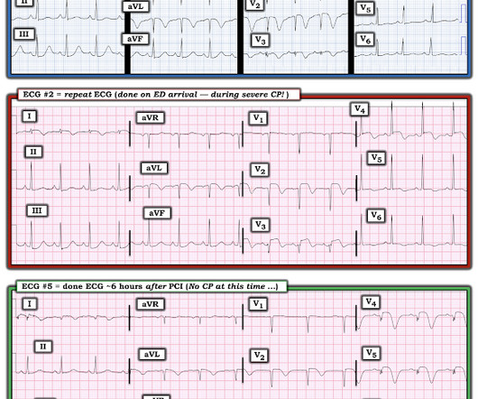
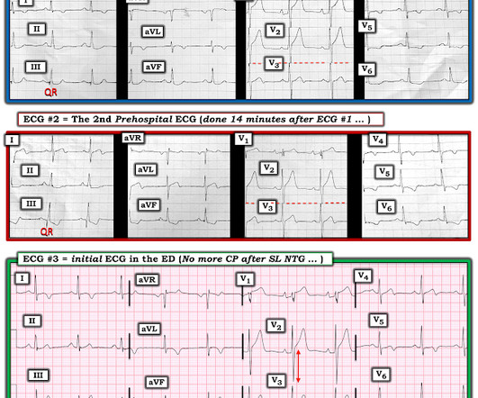
A 60-something awoke with 10/10 crushing chestpain. The angiogram showed an open artery with 95% stenosis and thrombosis and it was stented. But the patient's chestpain continues and so you order a 2nd ECG (ECG 2 here). He walked in to triage. I would expect that a stent would be placed.
Previous medical interventions included a spectrum of procedures, including catheter-directed thrombectomy for popliteal artery aneurysms with thrombosis, vascular bypass grafting for cerebral-anterior communicating artery aneurysms and arch replacement and stent implantation for aortic dissecting aneurysms.
The patient had experienced one month of chestpains, coughing and hemoptysis symptoms. Ultrasound showed no thrombosis in the veins of both lower limbs. The patient's father and grandfather have a history of lower limb venous thrombosis. The patient was diagnosed with acute pulmonary embolism and pneumonia.
5 We are aware that the current consensus is that the propensity for plaques to rupture is independent of plaque size; however, in our opinion, the hypothesis that small atherosclerotic plaques are the most likely to rupture, with resulting occlusive thrombosis, is unproven. These are typical findings at sites of plaque rupture.
A 70-something female with no previous cardiac history presented with acute chestpain. She awoke from sleep last night around 4:45 AM (3 hours prior to arrival) with pain that originated in her mid back. She stated the pain was achy/crampy. Over the course of the next hour, this pain turned into a pressure in her chest.
There was no chestpain. V1 and V2 are probably placed too high on the chest given close morphological similarity to aVR. Sudden narrowing of a coronary artery due to ACS (plaque rupture with thrombosis and/or downstream showering of platelet-fibrin aggregates). The fall was not a mechanical etiology. Type I ischemia.
In the last post, we saw how important old ECGs are in assessing the current ECG in a patient without atypical presentation (in this previous case, the patient had no chestpain, and the apparent inferior OMI did not have reciprocal ST depression in lead aVL). She has no chest symptoms. What is the diagnosis?
A man in his 70s with past medical history of hypertension, dyslipidemia, CAD s/p left circumflex stent 2 years prior presented to the ED with worsening intermittent exertional chestpain relieved by rest. This episode of chestpain began 3 hours ago and was persistent even at rest. Troponin was ordered. Am J Emerg Med.
This was a male in his 50's with a history of hypertension and possible diabetes mellitus who presented to the emergency department with a history of squeezing chestpain, lasting 5 minutes at a time, with several episodes over the past couple of months. Plan was for admission for chestpain workup. Jernberg T, et al.
This patient, who is a mid 60s female with a history of hypertension, hyperlipidemia and GERD, called 911 because of chestpain. A mid 60s woman with history of hypertension, hyperlipidemia, and GERD called 911 for chestpain. It is also NOT the clinical scenario of takotsubo (a week of intermittent chestpain).
Written by Pendell Meyers and Peter Brooks MD A man in his 30s with no known past medical history was reported to suddenly experience chestpain and shortness of breath at home in front of his family. Chestpain, SOB, Precordial T-wave inversions, and positive troponin. What is the Diagnosis? Now another, with ultrasound.
No chestpain. His inpatient clinicians did not think that an urgent angiogram was warranted given that he was chestpain free, his EKG appeared nondiagnostic, and serial troponins were not elevating beyond 2 ug/L. Patients on dialysis often do not have chestpain in the setting of acute MI. Why is this?
Case An elderly patient had acute chestpain and 911 was called. For this analysis, ACO was defined as angiographic evidence of coronary thrombosis with peak cardiac troponin-I (cTn-I) at least 10 ng/mL or cTn-T ≥ 1 ng/ mL. It is important for cardiologists to realize that a paramedic may see something they do not.
Written by Willy Frick A 40 year old woman was at home cooking when she developed chestpain. Furthermore, the operator worried about the patient's adherence to dual antiplatelet therapy, in which case she would be at risk for catastrophic stent thrombosis. She took an oxycodone and called EMS. I sent this to Drs.
link] A 62 year old man with a history of hypertension, type 2 diabetes mellitus, and carotid artery stenosis called 911 at 9:30 in the morning with complaint of chestpain. He described it as "10/10" intensity, radiating across his chest from right to left. This is written by Willy Frick, an amazing cardiology fellow in St.
But the symptoms returned with similar pattern – provoked by exertion, and alleviated with rest; except that on each occasion the chestpain was a little more intense, and the needed recovery period was longer in duration. Then, she attempted to reengage the activities at hand, and initially tolerated this well. Severe Hypoxia b.
The best course is to wait until the anatomy is defined by angio, then if proceeding to PCI, add Cangrelor (an IV P2Y12 inhibitor) I sent the ECG and clinical information of a 90-year old with chestpain to Dr. McLaren. His response: “subendocardial ischemia.
Cardiology felt her chestpain to be, most likely, the result of coronary supply-demand mismatch in the context of HCM endothelial remodeling (i.e. Below are two examples of this. Type II MI), however decided to pursue coronary angiogram out of an abundance of caution. References Naidu, S. American College of Cardiology.
He denied chestpain or dyspnea throughout. Nevertheless, I don't think a thrombosis related type I MI was ruled out here simply because the patient refused further evaluation. No previous study for comparison. Clinical Course: - He had no events on cardiac monitoring overnight. -
Despite standardized coronary intervention and anticoagulant/antiplatelet therapy, the patient reported intermittent chest discomfort with persistently elevated cardiac troponin and d-dimer levels 20 days after initial treatment. Repeat coronary angiography confirmed recurrent thrombosis in the right coronary artery.
This study reports a rare case of concurrent AMI and pulmonary thromboembolism in a patient diagnosed with pancreatic cancer.Case presentationA 70-year-old woman presented with acute chestpain and ST-segment elevation myocardial infarction, prompting immediate percutaneous coronary intervention (PCI) with the deployment of a drug-eluting stent.
This was texted to me by a paramedic while I was out running one day: "54 yo male chestpain started at 1pm. Here it is: Obvious inferior OMI, and now the STE in V1 is huge, with huge hyperacute T-waves of Right ventricular OMI The cath lab was re-activated: Angiogram: 100% occlusion mid-RCA occlusion (in-stent thrombosis).
He denied chestpain. This was most likely acute thrombosis of a coronary artery resulting in OMI: The ECG changes were attributed to hyperkalemia. It is correct that he did not have chestpain, but we must remember that fully 1/3 of full blown STEMI do not present with chestpain. The dye don't lie".except
Angiography was technically challenging as the patient was receiving CPR, but the cardiologist suspected acute stent thrombosis and initiated cangrelor, although no repeat angiography was able to be obtained. He had no chestpain, dyspnea, or any other anginal equivalent, and his vital signs were normal.
We organize all of the trending information in your field so you don't have to. Join thousands of users and stay up to date on the latest articles your peers are reading.
You know about us, now we want to get to know you!
Let's personalize your content
Let's get even more personalized
We recognize your account from another site in our network, please click 'Send Email' below to continue with verifying your account and setting a password.


Let's personalize your content