This site uses cookies to improve your experience. To help us insure we adhere to various privacy regulations, please select your country/region of residence. If you do not select a country, we will assume you are from the United States. Select your Cookie Settings or view our Privacy Policy and Terms of Use.
Cookie Settings
Cookies and similar technologies are used on this website for proper function of the website, for tracking performance analytics and for marketing purposes. We and some of our third-party providers may use cookie data for various purposes. Please review the cookie settings below and choose your preference.
Used for the proper function of the website
Used for monitoring website traffic and interactions
Cookie Settings
Cookies and similar technologies are used on this website for proper function of the website, for tracking performance analytics and for marketing purposes. We and some of our third-party providers may use cookie data for various purposes. Please review the cookie settings below and choose your preference.
Strictly Necessary: Used for the proper function of the website
Performance/Analytics: Used for monitoring website traffic and interactions
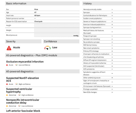
Written by Pendell Meyers Two patients with acute chestpain. Patient 1: Patient 2: Patient 1: A man in his 40s with minimal medical history presented with acute chestpain radiating to his R shoulder. Two patients with chestpain. Do either, neither, or both have OMI and need reperfusion?
The patient presented to an outside hospital An 80yo female per triage “patient presents with chestpain, also hurts to breathe” PMH: CAD, s/p stent placement, CHF, atrial fibrillation, pacemaker (placed 1 month earlier), LBBB. HPI: Abrupt onset of substernal chestpain associated with nausea/vomiting 30 min PTA.
A 50-something male with hypertension and 20- to 40-year smoking history presented with 1 week of stuttering chestpain that is worse with exertion, which takes many minutes to resolve after resting and never occurs at rest. At times the pain does go to his left neck. It is a ssociated with mild dyspnea on exertion.
Written by Jesse McLaren, comments by Smith A 55 year old with a history of NSTEMI presented with two hours of exertional chestpain, with normal vitals. Old ‘NSTEMI’ A history of coronary artery disease and a stent to the same territory further increases pre-test likelihood of acute coronary occlusion, including in-stent thrombosis.
A 40-something with severe diabetes on dialysis and with known coronary disease presented with acute crushing chestpain. 2 years prior he had an angiogram which showed 90% proximal stenosis of the circumflex. The pain did not resolve with NTG, and so he went to emergent angiography: 1. Left main: no obvious stenosis.
A 40-something male presented by ambulance with one hour of chestpain that was improving after sublingual nitroglycerine and 325 mg of aspirin, chewed. Here is the angiogram: --Culprit is 100% stenosis in the proximal RCA. (It It must have re-occluded between the ED and the cath lab) --Lesion was stented.
A 63 year old man with a history of hypertension, hyperlipidemia, prediabetes, and a family history of CAD developed chestpain, shortness of breath, and diaphoresis after consuming a large meal at noon. He called EMS, who arrived on scene about two hours after the onset of pain to find him hypertensive at 220 systolic.
The 50-something patient with history of coronary stenting and slightly reduced LV ejection fraction. In the setting of prior stenting and reduced left ventricular ejection fraction, would pursue a heart team revascularization approach Syntax score 28.5, Pericarditis would be even more unlikely in someone without chestpain.
A middle aged male presented at midnight after 14 hours of constant, severe substernal chestpain, radiating to his throat and to bilateral jaws, and associated with diaphoresis. The pain was not positional, pleuritic, or reproducible. It was not relieved by anything. He had no previous medical history. Is it STEMI or NonSTEMI?
Sent by Anonymous, written by Pendell Meyers A man in his 60s with history of CAD and 2 prior stents presented to the ED complaining of acute heavy substernal chestpain that began while eating breakfast about an hour ago, and had been persistent since then, despite EMS administering aspirin and nitroglycerin. Pre-intervention.
He had suffered a couple bouts of typical chestpain in the last 24 hours. This ECG (ECG #3) was recorded immediately after the last episode of pain spontaneously resolved. The pain had lasted about one hour. Case A 40-something male presented to triage. There are classic Wellens' waves in V2-V5. Am Heart J.
A 60-something awoke with 10/10 crushing chestpain. I would expect that a stent would be placed. The angiogram showed an open artery with 95% stenosis and thrombosis and it was stented. But the patient's chestpain continues and so you order a 2nd ECG (ECG 2 here). He walked in to triage.
Sent by anonymous A man in his 40s with no previous heart disease presented within 30 minutes of onset of acute chestpain that started while exercising. Angiogram findings included: 95% mid RCA stenosis with occluded distal right PDA secondary to thrombus (peristent OMI). Chestpain and a computer ‘normal’ ECG.
The patient's chestpain had resolved by the time of the ECG 2. But it does prove that the patient has coronary disease and makes the probability that his chestpain is due to ACS very very high. Angiogram: Widely patent RCA and LAD stents. Therefore, no stent was placed. (No There are well-formed Q-waves 3.
Here I annotate it: This shows 100% occluded circumflex (red arrow) and a 90% stenosis of the LAD (Yellow arrow). The LAD was thought to be not thrombotic, but a chronic tight stenosis. My THOUGHTS on ECG #1: We are told that the patient in today’s case had an episode of severe chestpain 3 nights prior to admission.
A prehospital “STEMI” activation was called on a 75 year old male ( Patient 1 ) with a history of hyperlipidemia and LAD and Cx OMI with stent placement. He arrived to the ED by helicopter at 1507, about three hours after the start of his chestpain while chopping wood around noon. It was stented. This was a large OMI.
On the second morning of his admission, he developed 10/10 chestpain and some diaphoresis after breakfast. The patient was given opiates which improved his chestpain to 7/10. The consulting cardiologist wrote in their note: “Could be cardiac chestpain. She is usually incredibly good at recognizing them!
The following day in the cath lab a borderline significant mid LAD stenosis was found. Decision was made to stent the lesion. There was never any severe chestpain. At cath the following day there was a borderline significant stenosis of the mid LAD with FFR 0,8. N-terminal-proBNP peaked at 18.000, then normalized.
A 60 yo with 2 previous inferior (RCA) STEMIs, stented, called 911 for one hour of chestpain. The first hs troponin I returned at 1100 ng/L Angiogram Lesion on 1st Obtuse Marginal : Proximal subsection = 90% stenosisStented. He had no h/o heart failure. DBT was 120 minutes, pretty good for a Non-STEMI OMI.
Submitted and written by Alex Bracey with edits by Pendell Meyers and Steve Smith Case A 50ish year old man with a history of CAD w/ prior LAD MI s/p LAD stenting presented to the ED with chestpain similar to his prior MI, but worse. The pain initially started the day prior to presentation. mV compared to 0.05-0.1
No patient with chestpain should be sent home without troponin testing. The red arrow shows a roughly 80% stenosis of the proximal LAD. The blue arrow shows another stenosis of the LAD distal to the first diagonal branch of about 99%. The green arrow shows a 95% stenosis of the ostium of the first diagonal branch.
This male in his 40's had been having intermittent chestpain for one week. He awoke from sleep with crushing central chestpain and called ems. EMS recorded a 12-lead, then gave 2 sublingual nitros with complete relief of pain. A stent was placed. Type B waves are deeper and symmetric. de Zwaan C.,
A 34 yo woman with a history of HTN, h/o SVT s/p ablation 2006, and 5 months post-partum presented with intermittent central chestpain and SOB. She had one episode of pain the previous night and two additional episodes early on morning the morning she presented. Deep breaths are painful and symptoms come and go.
It means either a percutaneous coronary intervention with a stent or CABG. You may be. But I am not.You need to undergo some re-vascularisation procedure. What do you mean by that Doctor ? Can I get my heart re-vascularised by drugs alone Doctor ? No we can’t. Hmmm , wait, we do have something called OMT/GDMT.
Case 1 A middle aged woman presented with acute chestpain and shortness of breath, unclear time since onset, and likely with episodic symptoms off and on throughout the day. Another lesion in the proximal LAD with 80% stenosis was stented as well. Normal RV, no valve stenosis or regurgitation. Angiogram @ 1830 (3.3
The patient said his chestpain was 4/10, down from 8/10 on presentation. The LAD has diffuse disease with a few areas of moderate stenosis but no flow-limiting lesions. On the combined basis of angiography and IVUS, this patient received stents to his mid RCA, proximal PDA, and OM. Repeat ECG at that time is shown.
This middle aged male with h/o GERD but also h/o stents presented to the ED with chestpain. The initial troponin I returned at 1500 ng/L and another ECG was recorded as the patient complained of 9/10 chestpain at 10 hours after the first Now the T-wave in III is fully upright, suggesting re-occlusion.
Case A 47 year old male called 911 for severe chestpain. It was opened and stented. A woman in her 60s with no prior history of CAD presented with 3 hours of sharp, centrally located chestpain with radiation to the anterior neck, with associated nausea. LM: No significant stenosis. Culprit, stented) 3.
This is the initial ED ECG of a 46 year old male with chestpain: The QTc was 420 ST Elevation at 60 ms after the J-point in lead V3 = 2.5 ng/ml) A 45 year old male called 911 for chestpain: The QTc was 400 ST Elevation at 60 ms after the J-point in lead V3 = 3.5 Angiogram showed a critical LAD thrombotic stenosis.
Written by Willy Frick A 40 year old woman was at home cooking when she developed chestpain. The cardiologist called this 20% stenosis. The operator documented thoughtful consideration of risks and benefits of stent placement. Unfortunately, a few hours later the patient complained of recurrent chestpain.
60-something with h/o MI and stents presented with chestpain radiating to the back and nausea/vomiting. The cath lab was activated: Result: Thrombotic 95% stenosis at the ostium of a small LPL2 with 70% stenosis at the LPL2/LPDA bifurcation in the distal/AV groove Cx Tubular 70% stenosis in the mid-circumflex. (In
link] A 62 year old man with a history of hypertension, type 2 diabetes mellitus, and carotid artery stenosis called 911 at 9:30 in the morning with complaint of chestpain. He described it as "10/10" intensity, radiating across his chest from right to left. Fortunately, that is exactly what happened.
There is evidence that de Winter's T-waves really represent a tiny trickle of blood through the thrombotic stenosis. Both were stented. New ST-depression (without LBBB or LVH) in aVL that cannot be blamed on an abnormal QRS is worrisome, and in the context of a patient with acute chestpain is almost certainly due to ischemia.
There is ventricular hypertrophy in the absence of abnormal loading conditions, such as aortic stenosis, or hypertension, for example – of which the most common variant is Asymmetric Septal Hypertrophy. A mid-LAD culprit lesion was identified and stented. Below are two examples of this.
She was treated medically for NonSTEMI, pending next day cath, which showed ulcerated plaque and a 60% thrombotic stenosis in the LAD distal to the first diagonal. It was stented. Echo may be normal ( especially if the patient no longer has chestpain ). Regional wall motion abnormality-distal septum and apex.
A 50-something with no previous cardiac history and no risk factors presented to the ED with acute chestpain (pressure) that radiated to the left arm. It was stented with good results. An ECG was immediately recorded: Computer read: Normal ECG What do you think? There is ST depression in V1-V3. Supplies a very large OM1.
Edits by Meyers and Smith A man in his 70s with PMH of hypertension, hyperlipidemia, type 2 diabetes, CVA, dual-chamber Medtronic pacemaker, presented to the ED for evaluation of acute chestpain. So the patient was taken for emergent cath, showing: Culprit artery: LAD (100% stenosis, TIMI 0) requiring thrombectomy and stent.
ChestPain – Benign Early Repol or OMI? Written by Destiny Folk, MD, Adam Engberg, MD, and Vitaliy Belyshev MD A man in his early 60s with a past medical history of hypertension, type 2 diabetes, obesity, and hyperlipidemia presented to the emergency department for evaluation of chestpain.
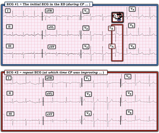
The patient’s chestpain spontaneously resolved before he was evaluated and has a repeat ECG obtained at 22:12 obtained shown below. In context, of course, it is clear that the patient is reperfusing, as pain has dissipated and the diagnostic findings of OMI have become more nonspecific. This ECG is more difficult.
He was at the gym when he had the onset of chestpain. Angiography revealed a very tight LAD stenosis with some flow (confirming the reperfusion that we see on the ECG). A stent was placed, and the patient had an excellent outcome with no wall motion abnormality. This patient is 38 years old with hyperlipidemia.
This means that at every age, the probability a man complaining of chestpain has significant underlying coronary disease as a cause of this chestpain is much higher than a woman complaining of chestpain. The data is overwhelming every way you can possibly look at it. years of age versus 59.0±8.4
Written by Pendell Meyers, edits by Steve Smith A man in his 60s with history of hypertension and MI 10 years ago, with PCI, presented to an outside hospital complaining of chestpain that started while mowing the lawn. The LAD lesion was acute and required 3 stents to restore flow. Here is his ECG on arrival: What do you think?
A middle-aged male with h/o CAD and stents presented with typical chest pressure. Case 3 : Male in 30's with chestpain, cough, and fever. A male in late middle age with a history of RCA stent 8 years prior complained of chestpain. This is a very common misread. What do you think? Called 911.
His comments/questions are inserted below the ECG: A 50-something woman presented with 3 days of intermittent chestpain that became worse on the day of presentation, with diaphoresis and radiation to the left arm, as well as abdominal pain. Everolimus-Eluting Stents or Bypass Surgery for Left Main Coronary Artery Disease.
We organize all of the trending information in your field so you don't have to. Join thousands of users and stay up to date on the latest articles your peers are reading.
You know about us, now we want to get to know you!
Let's personalize your content
Let's get even more personalized
We recognize your account from another site in our network, please click 'Send Email' below to continue with verifying your account and setting a password.


Let's personalize your content