This site uses cookies to improve your experience. To help us insure we adhere to various privacy regulations, please select your country/region of residence. If you do not select a country, we will assume you are from the United States. Select your Cookie Settings or view our Privacy Policy and Terms of Use.
Cookie Settings
Cookies and similar technologies are used on this website for proper function of the website, for tracking performance analytics and for marketing purposes. We and some of our third-party providers may use cookie data for various purposes. Please review the cookie settings below and choose your preference.
Used for the proper function of the website
Used for monitoring website traffic and interactions
Cookie Settings
Cookies and similar technologies are used on this website for proper function of the website, for tracking performance analytics and for marketing purposes. We and some of our third-party providers may use cookie data for various purposes. Please review the cookie settings below and choose your preference.
Strictly Necessary: Used for the proper function of the website
Performance/Analytics: Used for monitoring website traffic and interactions
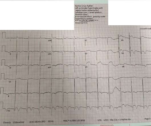
This is a previously healthy male teenager who was awoken by chestpain. The pain is described as located in the midsternal area, radiating to the right arm, described as 8-9/10 and worse with deep inspirations. In the evening, he became diaphoretic and complained of 9/10 continuous chestpain.
He denied chestpain or shortness of breath. In the clinical context of weakness and fever, without chestpain or shortness of breath, the likelihood of Brugada pattern is obviously much higher. Pediatric and elderly patients were more predisposed to developing an arrhythmic event in the setting of fever [7].
There was no chestpain. Extensive conduction system abnormalities can have various causes (ischemia, genetic, infectious, amyloid, etc). This was written by Magnus Nossen The patient is a female in her 50s. She presented with a one week hx of «dizziness» and weakness. She was feeling fine prior to the last seven days.
It was from a patient with chestpain: Note the obvious Brugada pattern. Pediatric and elderly patients were more predisposed to developing an arrhythmic event in the setting of fever [7]. The elevated troponin was attributed to either type 2 MI or to non-MI acute myocardial injury. There is no further workup at this time.
She did not even need to ask in this case, because even if the patient presented with chestpain, she would call it NEGATIVE. This ST-T wave pattern in lead V5 is not seen in other leads, as would be expected if this was truly a change of acute ischemia. What about the R = S Phenomenon in the Inferior Leads?
We organize all of the trending information in your field so you don't have to. Join thousands of users and stay up to date on the latest articles your peers are reading.
You know about us, now we want to get to know you!
Let's personalize your content
Let's get even more personalized
We recognize your account from another site in our network, please click 'Send Email' below to continue with verifying your account and setting a password.


Let's personalize your content