This site uses cookies to improve your experience. To help us insure we adhere to various privacy regulations, please select your country/region of residence. If you do not select a country, we will assume you are from the United States. Select your Cookie Settings or view our Privacy Policy and Terms of Use.
Cookie Settings
Cookies and similar technologies are used on this website for proper function of the website, for tracking performance analytics and for marketing purposes. We and some of our third-party providers may use cookie data for various purposes. Please review the cookie settings below and choose your preference.
Used for the proper function of the website
Used for monitoring website traffic and interactions
Cookie Settings
Cookies and similar technologies are used on this website for proper function of the website, for tracking performance analytics and for marketing purposes. We and some of our third-party providers may use cookie data for various purposes. Please review the cookie settings below and choose your preference.
Strictly Necessary: Used for the proper function of the website
Performance/Analytics: Used for monitoring website traffic and interactions
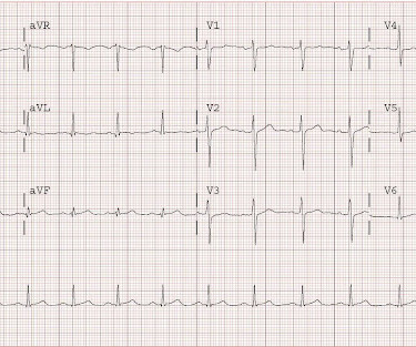
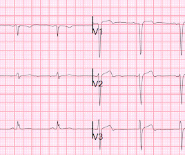
Written by Magnus Nossen with Edits by Grauer and Smith The ECGs in today’s case are from 3 different patients all presenting with new-onset CP ( ChestPain ). Elevated troponins prompted an echocardiogram — which revealed an apical wall motion abnormality (WMA). Patient #1 in today's post did not get expert ECG interpretation.
Given the presentation, the cardiologist stented the vessel and the patient returned to the ICU for ongoing critical care. Echocardiogram showed LVEF 66% with normal wall motion and normal diastolic function. He did not remember whether he had experienced any chestpain. Two subsequent troponins were down trending.
Case written and submitted by Ryan Barnicle MD, with edits by Pendell Meyers While vacationing on one of the islands off the northeast coast, a healthy 70ish year old male presented to the island health center for an evaluation of chestpain. The chestpain started about one hour prior to arrival while bike riding.
A 60-something man presented by EMS with 5 hours of fairly typical sounding substernal chestpain. EMS gave 324 mg aspirin and 3 sublingual NTG, which the patient stated reduced the substernal chestpain from an 8/10 to 4/10. Pain better still. What do you think the echocardiogram shows? NTG drip started.
The 50-something patient with history of coronary stenting and slightly reduced LV ejection fraction. In the setting of prior stenting and reduced left ventricular ejection fraction, would pursue a heart team revascularization approach Syntax score 28.5, Pericarditis would be even more unlikely in someone without chestpain.
While in the ED, patient developed acute dyspnea while at rest, initially not associated with chestpain. He later developed mild continuous chestpain, that he describes as the sensation of someone standing on his chest. See this post: What do you think the echocardiogram shows in this case?
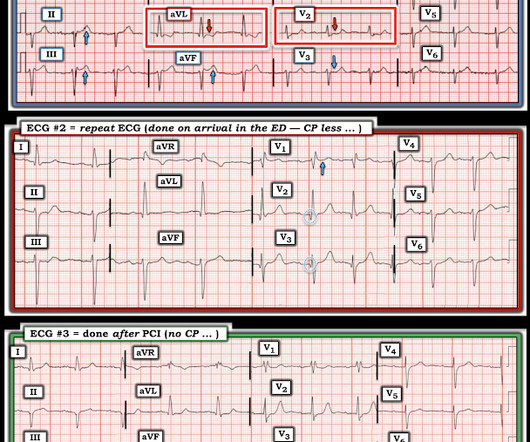
The patient’s chestpain spontaneously resolved before he was evaluated and has a repeat ECG obtained at 22:12 obtained shown below. In context, of course, it is clear that the patient is reperfusing, as pain has dissipated and the diagnostic findings of OMI have become more nonspecific. This ECG is more difficult.
A male in his 40's who had been discharged 6 hours prior after stenting of an inferoposterior STEMI had sudden severe SOB at home 2 hours prior to calling 911. He had no chestpain. Medications were aspirin, clopidogrel, metoprolol, and simvastatin. He was in acute distress from pulmonary edema, with a BP of 180/110, pulse 110.
Previous medical interventions included a spectrum of procedures, including catheter-directed thrombectomy for popliteal artery aneurysms with thrombosis, vascular bypass grafting for cerebral-anterior communicating artery aneurysms and arch replacement and stent implantation for aortic dissecting aneurysms.
Submitted by Benjamin Garbus, MD with edits by Bracey, Meyers, and Smith A man in his early 30s presented to the ED with chestpain described as an “explosion" of left chest pressure. Today’s pain lasted around 20 mins, but was severe enough that the patient called EMS. Triage EKG: What do you think? Do NOT use them.
Sent by anonymous A man in his 40s with no previous heart disease presented within 30 minutes of onset of acute chestpain that started while exercising. Successful drug-eluting stent placement opening up 95% mid RCA stenosis to 0% residual Nonobstructive left system disease. Chestpain and a computer ‘normal’ ECG.
An echocardiogram showed newly reduced left ventricular ejection fraction of 30-35%. He had minimal in-stent restenosis on angiography but had only minimal cardiac enzyme elevation and did not have antecedent chestpain before either of his events.
A prehospital “STEMI” activation was called on a 75 year old male ( Patient 1 ) with a history of hyperlipidemia and LAD and Cx OMI with stent placement. He arrived to the ED by helicopter at 1507, about three hours after the start of his chestpain while chopping wood around noon. It was stented. This was a large OMI.
On the second morning of his admission, he developed 10/10 chestpain and some diaphoresis after breakfast. The patient was given opiates which improved his chestpain to 7/10. The consulting cardiologist wrote in their note: “Could be cardiac chestpain. She is usually incredibly good at recognizing them!
See this case: Persistent ChestPain, an Elevated Troponin, and a Normal ECG. This is different from nitroglycerin which produces vasodilation and can improve by pain improving myocardial perfusion. Here is the angiogram after stent placement. See this case: A man his 50s with chestpain. At midnight.
A 60 yo with 2 previous inferior (RCA) STEMIs, stented, called 911 for one hour of chestpain. The first hs troponin I returned at 1100 ng/L Angiogram Lesion on 1st Obtuse Marginal : Proximal subsection = 90% stenosis Stented. He had no h/o heart failure. Pre procedure TIMI III flow was noted.
Submitted and written by Alex Bracey with edits by Pendell Meyers and Steve Smith Case A 50ish year old man with a history of CAD w/ prior LAD MI s/p LAD stenting presented to the ED with chestpain similar to his prior MI, but worse. The pain initially started the day prior to presentation. mV compared to 0.05-0.1
This male in his 40's had been having intermittent chestpain for one week. He awoke from sleep with crushing central chestpain and called ems. EMS recorded a 12-lead, then gave 2 sublingual nitros with complete relief of pain. A stent was placed. Type B waves are deeper and symmetric.
He continued to have worsening pain and diaphoresis, and associated left arm pain down to the fingers. reports MI in 2001 with a stent placed in the "marginal" artery. Pain is similar, but associated with less SOB. A stat echocardiogram would have helped to make this diagnosis and facilitate timely reperfusion.
He reported typical chestpain since 4H AM and arrived at our ED at 10h with ongoing chestpain. Successful primary angioplasty of the mid-circumflex artery towards the main marginal branch with the implantation of a drug-eluting stent. You must record frequent serial ECGs for patients with chestpain.
No patient with chestpain should be sent home without troponin testing. An echocardiogram showed severely reduced global systolic function with an EF of 20-25% and an LV apical thrombus. All three lesions had TIMI 2 flow prior to stenting. An echocardiogram showed an EF of 20-25%.
Written by Willy Frick A 40 year old woman was at home cooking when she developed chestpain. The patient was thought to have low likelihood of ACS, and cardiology recommended repeat troponin, urine drug testing, and echocardiogram. Bedside echocardiogram showed hypokinesis of the mid to distal anterior wall and apex.
This middle aged male with h/o GERD but also h/o stents presented to the ED with chestpain. The initial troponin I returned at 1500 ng/L and another ECG was recorded as the patient complained of 9/10 chestpain at 10 hours after the first Now the T-wave in III is fully upright, suggesting re-occlusion.
A middle aged male with no h/o CAD presented with one week of crescendo exertional angina, and had chestpain at the time of the first ECG: Here is the patient's previous ECG: Here is the patient's presenting ED ECG: There is isolated ST depression in precordial leads, deeper in V2 - V4 than in V5 or V6. There is no ST elevation.
Case 1 A middle aged woman presented with acute chestpain and shortness of breath, unclear time since onset, and likely with episodic symptoms off and on throughout the day. Another lesion in the proximal LAD with 80% stenosis was stented as well. Culprit lesion was reduced to 0% and stented. Additional case by Smith.
This was a male in his 50's with a history of hypertension and possible diabetes mellitus who presented to the emergency department with a history of squeezing chestpain, lasting 5 minutes at a time, with several episodes over the past couple of months. Plan was for admission for chestpain workup. It was stented.
link] A 62 year old man with a history of hypertension, type 2 diabetes mellitus, and carotid artery stenosis called 911 at 9:30 in the morning with complaint of chestpain. He described it as "10/10" intensity, radiating across his chest from right to left. His echocardiogram showed normal wall motion.
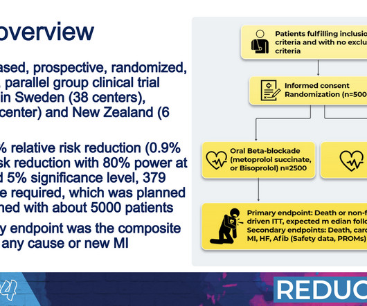
“At that time, the damage to the heart muscle was greater than we see today, and we didn’t have the possibility to revascularize patients with percutaneous coronary intervention and stents like we do today,” Yndigegn said. Half were randomly assigned to receive long-term beta blocker medications and the rest did not take beta blockers.
His medical history is unremarkable except a similar pain occurred 4-5 times in the previous 3 months with less intensity, short duration, unrelated to exertion. He visited an outpatient clinic for it and an echocardiogram and exercise stress test was normal. The lesion was successfully stented. Aspirated thrombotic material.
A middle-aged male called 911 for chestpain. Here was the first prehospital ECG with pain at 5/10: Computerized QTc is 418 ms. Formula value is now down to a very low value of 19.352 A 90% thrombotic LAD lesion was found and stented. There is nondiagnostic ST elevation in V1-V4. Most ST elevation is resolved.
No chestpain. I think a good start would be a posterior EKG and a high quality contrast echocardiogram read by an expert. His inpatient clinicians did not think that an urgent angiogram was warranted given that he was chestpain free, his EKG appeared nondiagnostic, and serial troponins were not elevating beyond 2 ug/L.
The lesion was stented. These are reperfusion T-waves (the same thing as Wellens' waves) Echocardiogram Regional wall motion abnormality-distal septum and apex. As emphasized by Dr. Smith — the best way to demonstrate acute ischemia is by identifying d ynamic S T- T wave c hanges in association with change in the nature of chestpain.
What do you think of this ECG in a patient with chestpain? Case history A middle-aged woman with a history of HTN, but no prior CAD, presented to the ED with chestpain. The pain had been mild and intermittent for 2 weeks, but had become more intense on the night of presentation. Is the ST elevation due to LVH?
A 40 something otherwise healthy man presented with substernal chestpain. They were stented. Formal Echocardiogram: The estimated left ventricular ejection fraction is 58 %. It had occurred once 3 days prior and resolved without any medical visit. What do you think? This ECG is DIAGNOSTIC of acute LAD Occlusion.
Case A 39-year-old male without prior medical history presents with chestpain that started 2 hours prior to presentation. He says that the pain intensity was 10/10 at home but now about 4/10. Despite the clinical stability and decreasing pain, this patient needs an immediate angiogram. Here are his publications.)
Written by Pendell Meyers An elderly female called EMS with acute chestpain. Rather, they tell you what has been going on in the last several hours: 1) Echocardiogram -- The myocardium remains stunned and hypokinetic after reperfusion. Her vitals were within normal limits, and here is her EMS ECG: What do you think?
Written by Willy Frick A man in his 60s with hypertension and prior stroke presented with three days of crushing chestpain. He reported intermittent chestpain for the last few months, but never lasting this long. The cardiology consultant notes that pain is "almost resolved." There is active infarction.
This is a very bold statement in a type 1 diabetic with very concerning sounding chestpain. The patient was treated with aspirin and a GI cocktail, which did not help the pain. Echocardiogram was finally performed five hours after the first diagnostic ECG. Echocardiogram showed LVEF 33% with akinesis of the lateral wall.
Written by Pendell Meyers, few edits by Smith A woman in her 70s was woken from sleep by midsternal chestpain radiating to left arm with nausea. Echocardiogram: EF 42% Moderate hypokinesis of the mid anterolateral and apical lateral myocardium Final diagnosis by the cardiologist was "STEMI" despite never meeting STEMI criteria.
Written by Pendell Meyers A man in his 60s presented with acute chestpain. Total proximal LAD occlusion was found and stented at angiography soon after the ECG above. Here is his triage ECG: What do you think? There is sinus rhythm with clear LVH. Here are some images: Next morning ECG: Reperfusion findings are clear.
We organize all of the trending information in your field so you don't have to. Join thousands of users and stay up to date on the latest articles your peers are reading.
You know about us, now we want to get to know you!
Let's personalize your content
Let's get even more personalized
We recognize your account from another site in our network, please click 'Send Email' below to continue with verifying your account and setting a password.


Let's personalize your content