This site uses cookies to improve your experience. To help us insure we adhere to various privacy regulations, please select your country/region of residence. If you do not select a country, we will assume you are from the United States. Select your Cookie Settings or view our Privacy Policy and Terms of Use.
Cookie Settings
Cookies and similar technologies are used on this website for proper function of the website, for tracking performance analytics and for marketing purposes. We and some of our third-party providers may use cookie data for various purposes. Please review the cookie settings below and choose your preference.
Used for the proper function of the website
Used for monitoring website traffic and interactions
Cookie Settings
Cookies and similar technologies are used on this website for proper function of the website, for tracking performance analytics and for marketing purposes. We and some of our third-party providers may use cookie data for various purposes. Please review the cookie settings below and choose your preference.
Strictly Necessary: Used for the proper function of the website
Performance/Analytics: Used for monitoring website traffic and interactions
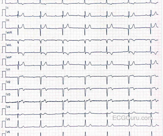
This is an interesting case for your students who want to delve into dysrhythmias with an eye on detail. I will start the discussion by admitting that I am not an expert of electrophysiology or complex dysrhythmias. I hope some of our dysrhythmia Gurus will delve into the rhythm and maybe even provide laddergrams.
This is an interesting case for your students who want to delve into dysrhythmias with an eye on detail. I will start the discussion by admitting that I am not an expert of electrophysiology or complex dysrhythmias. I hope some of our dysrhythmia Gurus will delve into the rhythm and maybe even provide laddergrams.
There was apparently no syncope and he had no bony injuries, but he did complain of left sided chestpain. His chest was tender. A Patient with Ischemic symptoms and a Biventricular Pacemaker A bedside cardiac ultrasound was normal. An ECG was recorded: Avinash was understandably confused by this ECG. IVCD type rhythm ??
This 60-something with h/o COPD and HFrEF (EF 25%) presented with SOB and chestpain. MAT has at least 3 distinct P-wave morphologies, but there is no single dominant pacemaker (i.e., The patient in this case presented with dyspnea and chestpain. Here is the ECG: What do you think?
Check : [vitals, SOB, ChestPain, Ultrasound] If the patient has Abdominal Pain, ChestPain, Dyspnea or Hypoxemia, Headache, Hypotension , then these should be considered the primary chief complaint (not syncope). Aortic Dissection, Valvular (especially Aortic Stenosis), Tamponade. Syncope with Exertion (EGSYS) 7.
We organize all of the trending information in your field so you don't have to. Join thousands of users and stay up to date on the latest articles your peers are reading.
You know about us, now we want to get to know you!
Let's personalize your content
Let's get even more personalized
We recognize your account from another site in our network, please click 'Send Email' below to continue with verifying your account and setting a password.


Let's personalize your content