This site uses cookies to improve your experience. To help us insure we adhere to various privacy regulations, please select your country/region of residence. If you do not select a country, we will assume you are from the United States. Select your Cookie Settings or view our Privacy Policy and Terms of Use.
Cookie Settings
Cookies and similar technologies are used on this website for proper function of the website, for tracking performance analytics and for marketing purposes. We and some of our third-party providers may use cookie data for various purposes. Please review the cookie settings below and choose your preference.
Used for the proper function of the website
Used for monitoring website traffic and interactions
Cookie Settings
Cookies and similar technologies are used on this website for proper function of the website, for tracking performance analytics and for marketing purposes. We and some of our third-party providers may use cookie data for various purposes. Please review the cookie settings below and choose your preference.
Strictly Necessary: Used for the proper function of the website
Performance/Analytics: Used for monitoring website traffic and interactions
This is an interesting case for your students who want to delve into dysrhythmias with an eye on detail. I will start the discussion by admitting that I am not an expert of electrophysiology or complex dysrhythmias. I hope some of our dysrhythmia Gurus will delve into the rhythm and maybe even provide laddergrams.
This is an interesting case for your students who want to delve into dysrhythmias with an eye on detail. I will start the discussion by admitting that I am not an expert of electrophysiology or complex dysrhythmias. I hope some of our dysrhythmia Gurus will delve into the rhythm and maybe even provide laddergrams.
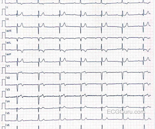
There was apparently no syncope and he had no bony injuries, but he did complain of left sided chestpain. His chest was tender. A bedside cardiac ultrasound was normal. An ECG was recorded: Avinash was understandably confused by this ECG. He wrote: "ECG 1 - shows wide ???IVCD IVCD type rhythm ?? What is it? What is the rhythm?
In that sense, the term dysrhythmia is preferable because it does literally translate as a disturbance in normal rhythm which is exactly what it is meant to describe. Any unsolicited disturbance of the rate or rhythm can be termed a dysrhythmia and result in the heart beating less efficiently but only for the duration of the dysrhythmia.
An elderly dialysis patient presented with chestpain. Relative contraindications to both include a known prolonged QT, especially if the dysrhythmia is believed to be caused by prolonged QT. She has poor LV function. Severely decreased LV function. Here is her ECG: Regular Wide Complex Tachycardia.
Written by Bobby Nicholson MD and Pendell Meyers A man in his 30s presented to the ED for evaluation of chestpain and palpitations. At this point, the patient had been symptomatic for almost 5 hours, appeared unwell with chestpain and diaphoresis. Thus, the patients rhythm is atrial fibrillation with WPW.
A 90 yo with a history of orthostatic hypotension had a near syncopal event followed by chestpain. Chestpain was resolved upon arrival in the ED. The second explanation (AIVR), whether as a reperfusion dysrhythmia or not, seems most likely. His previous ECG was normal. What is it? Answer below.
He denied chestpain or shortness of breath. In the clinical context of weakness and fever, without chestpain or shortness of breath, the likelihood of Brugada pattern is obviously much higher. There were no dysrhythmias on cardiac monitor during observation. See below for PM Cardio digitized version of this.
This was written by Magnus Nossen, from Norway, with comments and additions by Smith A 50 something smoker with no previous medical hx contacted EMS due to acute onset chestpain. Upon EMS arrival the patient appeared acutely ill and complained of chestpain. Is it sinus or is it a supraventricular dysrhythmia?
ECG of pneumopericardium and probable myocardial contusion shows typical pericarditis Male in 30's, 2 days after Motor Vehicle Collsion, complains of ChestPain and Dyspnea Head On Motor Vehicle Collision. Gunshot wound to the chest with ST Elevation Would your radiologist make this diagnosis, or should you record an ECG in trauma?
I also believe that we physicians and medics are eager to treat dysrhythmias, and we want to see them even when they are not there. Dilated pupils and hypertension are a strong clue to sympathetic overload, but don't forget anticholinergic syndromes, including tricyclics! Marcus, G. Harvard Medical School, Boston, Massachusetts, USA.
He had concurrent sharp substernal chestpain that resolved, but palpitations continued. Over past 3 months, he has had similar intermittent episodes of sharp chestpain while running, but none at rest. If you don't know what the dysrhythmia is, then try procainamide. What to do now?
Written by Pendell Meyers and Peter Brooks MD A man in his 30s with no known past medical history was reported to suddenly experience chestpain and shortness of breath at home in front of his family. Chestpain, SOB, Precordial T-wave inversions, and positive troponin. What is the Diagnosis? Now another, with ultrasound.
Now you have ECG and troponin evidence of ischemia, AND ventricular dysrhythmia, which means this is NOT a stable ACS. While ST coving in V1 is not necessarily abnormal — the presence of ST elevation in association with ST-T wave abnormalities in V2,V3 in a patient with chestpain is clearly cause for concern.
Opinions vary widely on the K level at which a patient must be admitted on a monitor because of the risk of ventricular dysrhythmias. My rationale is that if the K is affecting the ECG, then it is affecting the electrical milieu and can result in serious dysrhythmias. Until some real data is available, my opinion is this: 1.
She reports that she is now unable to vagal out of her palpitations and is having shortness of breath and dull chestpain. But adenosine only lasts for seconds, and if the dysrhythmia recurs, then the adenosine is gone. Prevent the initiation of the dysrhythmia -- this can be done with a beta blocker by prenenting PACS 2.
A late middle-aged man presented with one hour of chestpain. Could the dysrhythmias have been prevented? Severe hypokalemia in the setting of STEMI or dysrhythmias is life-threatening and needs very rapid treatment. Most recent echo showed EF of 60%. He also had a history of chronic kidney disease, stage III.
This 60-something with h/o COPD and HFrEF (EF 25%) presented with SOB and chestpain. Atrial dysrhythmias, and atrial fi brillation in particular, are frequently misdiagnosed by computer algorithms and then by the physician who overreads them. The patient in this case presented with dyspnea and chestpain.
He was admitted for monitoring, as his risk of a ventricular dysrhythmia as cause of the syncope is high ( very high due to HFrEF and ischemic cardiomyopathy ). He denied chestpain or dyspnea throughout. No previous study for comparison. Clinical Course: - He had no events on cardiac monitoring overnight. -
It was from a patient with chestpain: Note the obvious Brugada pattern. The elevated troponin was attributed to either type 2 MI or to non-MI acute myocardial injury. There is no further workup at this time. Smith: Here is a case that was just texted to me today from a former resident. This patient ruled out for MI.
Inferior MI results in scar tissue which is a likely source of a re-entrant ventricular dysrhythmia. Here is the post-cardioversion ECG: There is sinus with RBBB There are inferior Q-waves suggesting old inferior MI. This would be the likely source of the VT.
It was edited by Smith CASE : A 52-year-old male with a past medical history of hypertension and COPD summoned EMS with complaints of chestpain, weakness and nausea. This was contributed by some folks at Wake Forest: Jason Stopyra, Shannon Mumma, Sean O'Rourke, and Brian Hiestand.
Check : [vitals, SOB, ChestPain, Ultrasound] If the patient has Abdominal Pain, ChestPain, Dyspnea or Hypoxemia, Headache, Hypotension , then these should be considered the primary chief complaint (not syncope). Aortic Dissection, Valvular (especially Aortic Stenosis), Tamponade.
This middle-aged man with no cardiac history but with significant history of methamphetamin and alcohol use presented with chestpain and SOB, worsening over days, with orthopnea. BP:143/99, Pulse 109, Temp 37.2 °C C (99 °F), Resp (!) 32, SpO2 95% On exam, he was tachypneic and had bibasilar crackles.
A 26 year old male presented with syncope and chestpain. No signs of OMI" The chestpain resolved after some time, and another ECG was recorded: The ST Elevation is nearly gone. He was admitted for monitoring and had no dysrhythmias. This appears to be an inferior OMI What do you think? She is very good.
Sinus tach is often misinterpreted as a dysrhythmia. With OMI, all you know is that your patient has some nonspecific chestpain, SOB, shoulder pain etc. They often have good ejection fraction and tolerate the dysrhythmia quite well. 2) PSVT with "aberrancy" (atypical RBBB+LAFB).
We organize all of the trending information in your field so you don't have to. Join thousands of users and stay up to date on the latest articles your peers are reading.
You know about us, now we want to get to know you!
Let's personalize your content
Let's get even more personalized
We recognize your account from another site in our network, please click 'Send Email' below to continue with verifying your account and setting a password.


Let's personalize your content