This site uses cookies to improve your experience. To help us insure we adhere to various privacy regulations, please select your country/region of residence. If you do not select a country, we will assume you are from the United States. Select your Cookie Settings or view our Privacy Policy and Terms of Use.
Cookie Settings
Cookies and similar technologies are used on this website for proper function of the website, for tracking performance analytics and for marketing purposes. We and some of our third-party providers may use cookie data for various purposes. Please review the cookie settings below and choose your preference.
Used for the proper function of the website
Used for monitoring website traffic and interactions
Cookie Settings
Cookies and similar technologies are used on this website for proper function of the website, for tracking performance analytics and for marketing purposes. We and some of our third-party providers may use cookie data for various purposes. Please review the cookie settings below and choose your preference.
Strictly Necessary: Used for the proper function of the website
Performance/Analytics: Used for monitoring website traffic and interactions
This was sent by anonymous The patient is a 55-year-old male who presented to the emergency department after approximately 3 to 4 days of intermittent central boring chestpain initially responsive to nitroglycerin, but is now more constant and not responsive to nitroglycerin. It is unknown when this pain recurred and became constant.
This is another case written by Pendell Meyers (who is helping to edit the blog and has many great recent posts) Case A 45 year old man was driving to work when he experienced acute onset sharp left sided chestpain with paresthesias of the left arm. A repeat ECG was recorded with pain 2/10: Not much change.
While in the hospital, he had witnessed ventricular fibrillation (VF) arrest for which he received external defibrillation. He had minimal in-stent restenosis on angiography but had only minimal cardiac enzyme elevation and did not have antecedent chestpain before either of his events.
Chris Mondie of the Newark Beth Israel Emergency Medicine Residency sent this case A 50-something man presented with acute chestpain. 100% proximal LAD successfully stented. Defibrillated out of v fib in the cath lab. Here is his ECG: As you can see, the computer called it completely normal What do you think?
The patient’s chestpain spontaneously resolved before he was evaluated and has a repeat ECG obtained at 22:12 obtained shown below. In context, of course, it is clear that the patient is reperfusing, as pain has dissipated and the diagnostic findings of OMI have become more nonspecific. This ECG is more difficult.
A 56 yo f with h/o HTN and hypercholesterolemia called EMS from home after onset of L chestpain radiating to the left arm. She was defibrillated successfully from ventricular fibrillation and developed a perfusing rhythm. Before EMS arrived, she had "seizure activity" and became unresponsive. She was intubated.
He was at the gym when he had the onset of chestpain. A stent was placed, and the patient had an excellent outcome with no wall motion abnormality. This patient is 38 years old with hyperlipidemia. There is a wide S-wave in V6. Thus, there is right bundle branch block, which should never (unlike Left BBB) have any ST elevation.
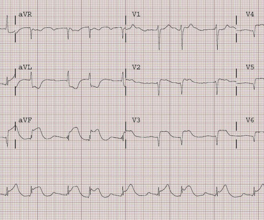
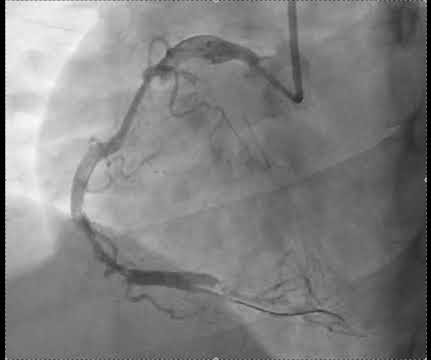
Written by Pendell Meyers, edits by Steve Smith A man in his 60s with history of hypertension and MI 10 years ago, with PCI, presented to an outside hospital complaining of chestpain that started while mowing the lawn. He was defibrillated immediately and had return of normal mental status.
A late middle-aged man presented with one hour of chestpain. At cath, he immediately had incessant Torsades de Pointes requiring defibrillation 7 times and requiring placement of a transvenous pacer for overdrive pacing at a rate of 80. This was stented. Most recent echo showed EF of 60%. He had recently had a NonSTEMI.
When a person experiences a heart attack or myocardial infarction, they may feel chestpain and other symptoms in different parts of their body. These issues can only be addressed in an ICCU (Intensive Coronary Care Unit) setting, where temporary pacemakers and defibrillators are available.
Written by Willy Frick A young woman with a history of paroxysmal nocturnal hemoglobinuria presented with acute substernal chestpain. The report describes heavy plaque in the proximal RCA by IVUS, but no lesions in the previously occluded RPL branch and no stent was deployed. Smith : The cath lab should be activated now!
Written by Willy Frick A woman in her 60s with very severe hyperlipidemia (LDL >200 mg/dL) presented with acute onset chestpain. She described the pain as moderate in severity, and said it had come and gone several times over the next few hours before ultimately resolving. Her symptoms began while getting off the bus.
After stent deployment, we often see improvement in the ST-T within seconds or minutes. Here is the final angiogram following placement of a stent in the ostial RCA. 2:04 PM, post stent deployment You can see that even after complete restoration of flow, the ECG still looks terrible, V most of all. SanzRuiz, R., Solis, J., &
Written by Pendell Meyers A man in his 60s presented with acute chestpain. Defibrillation was performed, and ROSC was achieved. Total proximal LAD occlusion was found and stented at angiography soon after the ECG above. Here is his triage ECG: What do you think? There is sinus rhythm with clear LVH.
We organize all of the trending information in your field so you don't have to. Join thousands of users and stay up to date on the latest articles your peers are reading.
You know about us, now we want to get to know you!
Let's personalize your content
Let's get even more personalized
We recognize your account from another site in our network, please click 'Send Email' below to continue with verifying your account and setting a password.


Let's personalize your content