This site uses cookies to improve your experience. To help us insure we adhere to various privacy regulations, please select your country/region of residence. If you do not select a country, we will assume you are from the United States. Select your Cookie Settings or view our Privacy Policy and Terms of Use.
Cookie Settings
Cookies and similar technologies are used on this website for proper function of the website, for tracking performance analytics and for marketing purposes. We and some of our third-party providers may use cookie data for various purposes. Please review the cookie settings below and choose your preference.
Used for the proper function of the website
Used for monitoring website traffic and interactions
Cookie Settings
Cookies and similar technologies are used on this website for proper function of the website, for tracking performance analytics and for marketing purposes. We and some of our third-party providers may use cookie data for various purposes. Please review the cookie settings below and choose your preference.
Strictly Necessary: Used for the proper function of the website
Performance/Analytics: Used for monitoring website traffic and interactions
A 50-something male had onset of chestpain 1 hour prior to ED arrival. Endorses some associated SOB, but denies back pain, fever, cough, chills, leg swelling, or other new symptoms. The patient was moved to the criticalcare area (stabilization room). Always get serial ECGs in a patient with acute chestpain.
This EKG was recorded as part of a standing order for criticalcare. Upon questioning patient, he denies having any chestpain or chest tightness of any sort. In the absence of chestpain and negative troponin , it appears less likely that he is having acute coronary syndrome though EKG appears concerning.
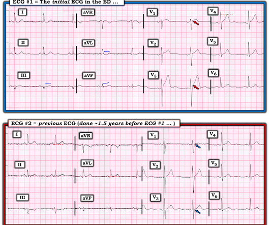
He reports significant chestpain at the base of his scapula on the right side along with new shortness of breath. Wellen's waves indicate that, when the patient was having chestpain, there was occlusion. See these casese (and I have many others): First ED ECG is Wellens' (pain free). A 70-something y.o.
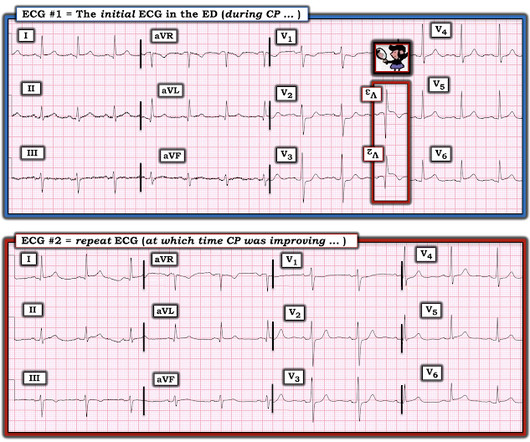
If you saw this ECG only knowing that it is an acute chestpain patient, what would be your interpretation? There was high suspicion of OMI, so patient was brought to criticalcare area and another ECG was recorded just 7 minutes later as the pain had diminished to 4/10. Left main: no significant stenosis.
This middle aged male with h/o GERD but also h/o stents presented to the ED with chestpain. The initial troponin I returned at 1500 ng/L and another ECG was recorded as the patient complained of 9/10 chestpain at 10 hours after the first Now the T-wave in III is fully upright, suggesting re-occlusion.
We knew only that the ECG belonged to a man in his 50s with chestpain and normal vitals. We brought the patient into one of our criticalcare rooms and immediately got more history while recording this repeat ECG: The STE in I has greatly diminished or entirely disappeared. No prior available. We activated the cath lab.
There was no chestpain. Later, I was working in the ED and a patient was moved from a regular room to the criticalcare area due to recurrent hypotension. The patient was now under my care. But today's patient had no chestpain. That condition is tricuspid stenosis, which is rare.
Colin is an emergency medicine resident beginning his criticalcare fellowship in the summer with a strong interest in the role of ECG in criticalcare and OMI. They had difficulty describing their symptoms, but complained of severe weakness, nausea, vomiting, headache, and chestpain. Edits by Willy Frick.
A 50-something with no previous cardiac history and no risk factors presented to the ED with acute chestpain (pressure) that radiated to the left arm. But even without these additional findings — the "Must Recognize" ECG pattern in this patient with new chestpain — is the unmistakeable shape of the ST depression in leads V2 and V3!
We organize all of the trending information in your field so you don't have to. Join thousands of users and stay up to date on the latest articles your peers are reading.
You know about us, now we want to get to know you!
Let's personalize your content
Let's get even more personalized
We recognize your account from another site in our network, please click 'Send Email' below to continue with verifying your account and setting a password.


Let's personalize your content