This site uses cookies to improve your experience. To help us insure we adhere to various privacy regulations, please select your country/region of residence. If you do not select a country, we will assume you are from the United States. Select your Cookie Settings or view our Privacy Policy and Terms of Use.
Cookie Settings
Cookies and similar technologies are used on this website for proper function of the website, for tracking performance analytics and for marketing purposes. We and some of our third-party providers may use cookie data for various purposes. Please review the cookie settings below and choose your preference.
Used for the proper function of the website
Used for monitoring website traffic and interactions
Cookie Settings
Cookies and similar technologies are used on this website for proper function of the website, for tracking performance analytics and for marketing purposes. We and some of our third-party providers may use cookie data for various purposes. Please review the cookie settings below and choose your preference.
Strictly Necessary: Used for the proper function of the website
Performance/Analytics: Used for monitoring website traffic and interactions
This was sent by anonymous The patient is a 55-year-old male who presented to the emergency department after approximately 3 to 4 days of intermittent central boring chestpain initially responsive to nitroglycerin, but is now more constant and not responsive to nitroglycerin. It is unknown when this pain recurred and became constant.
A 50-something male had onset of chestpain 1 hour prior to ED arrival. Endorses some associated SOB, but denies back pain, fever, cough, chills, leg swelling, or other new symptoms. The patient was moved to the criticalcare area (stabilization room). Always get serial ECGs in a patient with acute chestpain.
I was working at triage when the medics brought this patient who is 65 yo and has had chestpain for 12 hours. I took the patient to the criticalcare area and questioned him more on the way. The pain had been intermittent until an hour before arrival, when he called 911. So I uncrumpled it: What do you think?
Given the presentation, the cardiologist stented the vessel and the patient returned to the ICU for ongoing criticalcare. He did not remember whether he had experienced any chestpain. At his family's request, he was transferred to a hospital closer to his home to continue care. He was admitted to cardiology.
Colin is an emergency medicine resident beginning his criticalcare fellowship in the summer with a strong interest in the role of ECG in criticalcare and OMI. They had difficulty describing their symptoms, but complained of severe weakness, nausea, vomiting, headache, and chestpain. Edits by Willy Frick.
This EKG was recorded as part of a standing order for criticalcare. Upon questioning patient, he denies having any chestpain or chest tightness of any sort. In the absence of chestpain and negative troponin , it appears less likely that he is having acute coronary syndrome though EKG appears concerning.
A 50-something with no previous cardiac history and no risk factors presented to the ED with acute chestpain (pressure) that radiated to the left arm. But even without these additional findings — the "Must Recognize" ECG pattern in this patient with new chestpain — is the unmistakeable shape of the ST depression in leads V2 and V3!
I activated the cath lab and brought her to the criticalcare area. I went back to screening patients and time flew by and I forgot to get the 15 minute follow up ECG, but then remembered and did get one at about 45 minutes: Computer interpretation: "Normal ECG" Veritas algorithm again What do you think?
He reports significant chestpain at the base of his scapula on the right side along with new shortness of breath. Wellen's waves indicate that, when the patient was having chestpain, there was occlusion. See these casese (and I have many others): First ED ECG is Wellens' (pain free). A 70-something y.o.
This 54 year old patient with a history of kidney transplant with poor transplant function had been vomiting all day when at 10 PM he developed severe substernal crushing chestpain. He presented to the Emergency Department with a blood pressure of 111/66 and a pulse of 117. He had this ECG recorded.
If you saw this ECG only knowing that it is an acute chestpain patient, what would be your interpretation? There was high suspicion of OMI, so patient was brought to criticalcare area and another ECG was recorded just 7 minutes later as the pain had diminished to 4/10. Suspicious but not diagnostic.
He was a 30-something with chestpain. A male in his 30's complained of sudden severe substernal chestpain. Shoulder pain after lifting a heavy box de Winter's -- they remained stable for 30 minutes with many ECGs. Here is one case of a patient I saw. But we find that this is unusual. They are too narrow.
This middle aged male with h/o GERD but also h/o stents presented to the ED with chestpain. The initial troponin I returned at 1500 ng/L and another ECG was recorded as the patient complained of 9/10 chestpain at 10 hours after the first Now the T-wave in III is fully upright, suggesting re-occlusion.
The recent change in guidelines from the ACC and AHA are now clear that cardiac CT is the Level 1A evidence recommended test for diagnosis of stable and acute chestpain,” said Chaim Lotan, MD, professor at Hadassah-Hebrew University, in a written statement from Arineta. Workflow improvements.
Submitted and written by Alex Bracey, with edits by Pendell Meyers and Steve Smith: I was walking through the criticalcare section of the ED when I overheard a discussion about the following ECG. The patient was given fentanyl initially for chestpain with minimal effect and then vomited which was followed by zofran and famotidine.
The patient was otherwise healthy, had no past history, and had never had chest discomfort before. I immediately activated the criticalcare team and walked the patient to the criticalcare area, our "Stabilization Room." There are relatively large T-waves in V4-V6. Let's record another one."
We knew only that the ECG belonged to a man in his 50s with chestpain and normal vitals. We brought the patient into one of our criticalcare rooms and immediately got more history while recording this repeat ECG: The STE in I has greatly diminished or entirely disappeared. No prior available.
He denied any chestpain or shortness of breath and stated he felt at his baseline yesterday prior to drug use. They recommended repeating his ECG and awaiting troponin since the patient did not have any chestpain. He complained of generalized weakness and left lower extremity numbness. What is it?
The patient had come to the ED for SOB, but without any chestpain. He was moved to the criticalcare area due to his EKG. Other cases of Pulse Tapping Artifact: Acute chestpain and a bizarre ECG Bizarre (Hyperacute??) Solution : repeat the ECG, but move the left leg electrode. His potassium returned at 1.3
There was no chestpain. Later, I was working in the ED and a patient was moved from a regular room to the criticalcare area due to recurrent hypotension. The patient was now under my care. But today's patient had no chestpain. In reviewing the case, I saw the ECG and recognized it as the same one.
Instead, he complained of left chest "itchiness". He was brought to the criticalcare area where these rhythms were seen on the monitor: Wide complex tachycardia with no apparent P-waves, and very irregular Consistent with atrial fibrillation with aberrancy A Regular wide complex tachycardia. LV Aneurysm?
She denied chestpain and denied feeling any palpitations, even during her triage ECG: What do you think? Despite otherwise normal vital signs, she was appropriately triaged to the criticalcare area of the ED.
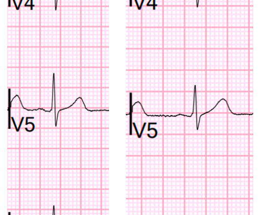
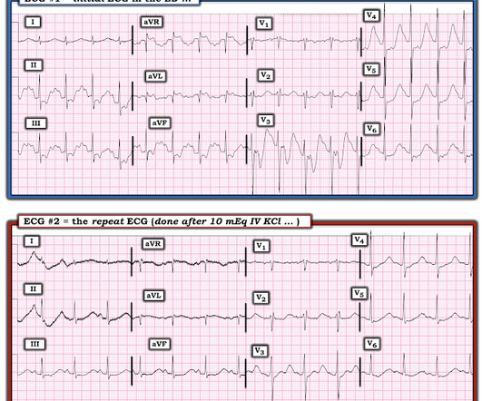
A late middle-aged man presented with one hour of chestpain. Crit Care Med. 1991 May;19(5):694-9 Objective: To evaluate the efficacy and safety of potassium replacement infusions in critically ill patients. Setting: Multidisciplinary criticalcare unit. Most recent echo showed EF of 60%.
A middle aged patient who was 3 weeks s/p STEMI came from cardiac rehab where he developed some chestpain, dyspnea and weakness on the treadmill. In the ED he had some continued chestpain and hypotension. Here was his ECG: There are inferior and lateral Q-waves with T-wave inversion in the corresponding leads.
Case 1: 20-something woman with chestpain Case 2: 50-something man with chestpain Case 1 A 20-something yo woman presented in the middle of the night with severe crushing chestpain. The blood pressure was 170/100 in the criticalcare area. Which patient needs a CT Scan? Denies SOB.
Dizziness is so unlikely to be OMI without an obvious ECG, that I am going to pretend that this patient presented with chestpain. So let's pretend this is acute chestpain. 20 cases with pseudonormalization Case continued The patient was moved to the criticalcare area, and cardiology was consulted.
1) Very high initial troponin of 45,000 ng/L 2) A full day of chestpain 3) Q-waves on the ECG, with some T-wave inversion Here is one frame of the CT scan which includes the heart: Can you spot the infarct? There is STE in V5-6. There are new Q-waves in aVL, V5-6. SUBACUTE) OMI, that would result in an undesirable delay.
Just a few weeks ago, I took care of a patient who had ostial RCA OMI (TIMI 0 at cath) and his only complaint was syncope! He had no chestpain, dyspnea, or any other anginal equivalent, and his vital signs were normal. Multidisciplinary criticalcare management of electrical storm. link] Jentzer, J. Kashou, A.
We organize all of the trending information in your field so you don't have to. Join thousands of users and stay up to date on the latest articles your peers are reading.
You know about us, now we want to get to know you!
Let's personalize your content
Let's get even more personalized
We recognize your account from another site in our network, please click 'Send Email' below to continue with verifying your account and setting a password.


Let's personalize your content