This site uses cookies to improve your experience. To help us insure we adhere to various privacy regulations, please select your country/region of residence. If you do not select a country, we will assume you are from the United States. Select your Cookie Settings or view our Privacy Policy and Terms of Use.
Cookie Settings
Cookies and similar technologies are used on this website for proper function of the website, for tracking performance analytics and for marketing purposes. We and some of our third-party providers may use cookie data for various purposes. Please review the cookie settings below and choose your preference.
Used for the proper function of the website
Used for monitoring website traffic and interactions
Cookie Settings
Cookies and similar technologies are used on this website for proper function of the website, for tracking performance analytics and for marketing purposes. We and some of our third-party providers may use cookie data for various purposes. Please review the cookie settings below and choose your preference.
Strictly Necessary: Used for the proper function of the website
Performance/Analytics: Used for monitoring website traffic and interactions
A 70-something female with no previous cardiac history presented with acute chestpain. She awoke from sleep last night around 4:45 AM (3 hours prior to arrival) with pain that originated in her mid back. She stated the pain was achy/crampy. Over the course of the next hour, this pain turned into a pressure in her chest.
A 63 year old man with a history of hypertension, hyperlipidemia, prediabetes, and a family history of CAD developed chestpain, shortness of breath, and diaphoresis after consuming a large meal at noon. He called EMS, who arrived on scene about two hours after the onset of pain to find him hypertensive at 220 systolic.
No prior exertional complaints of chestpain, dizziness, lightheadedness, or undue shortness of breath. He denied headache or neck pain associated with exertion. I sent this ECG to Dr. Smith, with the only information that it is a 17 year old with chestpain. 24 yo woman with chestpain: Is this STEMI?
Written by Pendell Meyers, edits by Smith and Grauer A man in his late 20s with history of asthma presented to the ED with a transient episode of chestpain and shortness of breath after finishing a 4-mile run. Ct coronaryangiogram showed normal coronary arteries. He typically runs 4 to 8 miles per day.
The medics stated he had been nauseated and diaphoretic, but he did not have any chestpain or SOB. And especially suspect Old MI when the patient gives a history of MI and has no chestpain or SOB. He had a prehospital ECG. He was ambulatory at the scene. He has a history of STEMI and heart failure.
Knowledge of this fundamental pillar of biology should drive how cardiologists approach men and women being evaluated for the presence of significant coronary disease. Atypical angina is classified as having any two of the three symptoms, and non-anginal pain any one of the three symptoms. versus 66.3%; P =0.004), older age (62.4±7.9
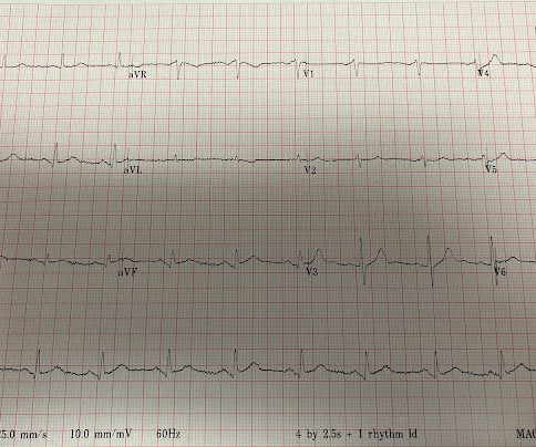
Hospital evaluation for this patient was negative for an acute coronary syndrome ( ie, CT coronaryangiogram was normal — troponin was not elevated — and Echo was negative, with no sign of pericardial effusion ). I interpreted the ECG in Figure-1 as follows: The rhythm is sinus at ~65/minute.
This was texted to me from a former resident, while working at a small rural hospital, with the statement: "I can’t convince myself of anything here, but he’s a 63-year-old guy with prior stents and a good story for ACS." Chestpain or discomfort) What do you think? The total duration of chestpain was 30-45 minutes.
But the symptoms returned with similar pattern – provoked by exertion, and alleviated with rest; except that on each occasion the chestpain was a little more intense, and the needed recovery period was longer in duration. It’s judicious, then, to arrange for coronaryangiogram. CoronaryAngiogram 1.
This worried the crew of potential acute coronary syndrome and STEMI was activated pre-hospital. When OMI is captured in this early phase, there exists the highest amount of salvageable myocardium and least likelihood of heart failure at hospital discharge. Smith comment : V5 and V6 are excessively discordant!!!!
He denied chestpain or dyspnea throughout. He was not interested in remaining hospitalized for further cardiac monitoring or inpatient EP consultation as he felt well, was convinced dehydration was the reason for his syncope, and he adamantly wanted to go home. Figure-1: The 2 ECGs in todays case ( See text ). See text ).
The best course is to wait until the anatomy is defined by angio, then if proceeding to PCI, add Cangrelor (an IV P2Y12 inhibitor) I sent the ECG and clinical information of a 90-year old with chestpain to Dr. McLaren. All electrocardiograms (ECGs) and coronaryangiograms were blindly analyzed by experienced cardiologists.
It was edited by Smith CASE : A 52-year-old male with a past medical history of hypertension and COPD summoned EMS with complaints of chestpain, weakness and nausea. The diagnostic coronaryangiogram identified only minimal coronary artery disease, but there was a severely calcified, ‘immobile’ aortic valve.
A 69 year old woman with a history of hypertension presented to the emergency department by EMS for evaluation of chestpain and shortness of breath. She awoke in the morning with sharp chestpain which worsened throughout the morning. As her pain worsened, so did her dyspnea. This was written by Hans Helseth.
We organize all of the trending information in your field so you don't have to. Join thousands of users and stay up to date on the latest articles your peers are reading.
You know about us, now we want to get to know you!
Let's personalize your content
Let's get even more personalized
We recognize your account from another site in our network, please click 'Send Email' below to continue with verifying your account and setting a password.


Let's personalize your content