This site uses cookies to improve your experience. To help us insure we adhere to various privacy regulations, please select your country/region of residence. If you do not select a country, we will assume you are from the United States. Select your Cookie Settings or view our Privacy Policy and Terms of Use.
Cookie Settings
Cookies and similar technologies are used on this website for proper function of the website, for tracking performance analytics and for marketing purposes. We and some of our third-party providers may use cookie data for various purposes. Please review the cookie settings below and choose your preference.
Used for the proper function of the website
Used for monitoring website traffic and interactions
Cookie Settings
Cookies and similar technologies are used on this website for proper function of the website, for tracking performance analytics and for marketing purposes. We and some of our third-party providers may use cookie data for various purposes. Please review the cookie settings below and choose your preference.
Strictly Necessary: Used for the proper function of the website
Performance/Analytics: Used for monitoring website traffic and interactions
Written by Colin Jenkins and Nhu-Nguyen Le with edits by Willy Frick and by Smith A 46-year-old male presented to the emergency department with 2 days of heavy substernal chestpain and nausea. The patient continued having chestpain. Circulation Research , 56 (2), 184–194. Circulation , 63 (2), 333–340.
They shocked him twice before return of spontaneous circulation. This was interpreted by the treating clinicians as not showing any evidence of ischemia. He did not remember whether he had experienced any chestpain. When EMS arrived the patient was in ventricular fibrillation. He was admitted to cardiology.
Another ECG was recorded after the nitroglycerine and now without pain: All findings are resolved. This confirms that the pain was ischemia and is now resovled. The history is concerning ( This patient was awakened from sleep by chestpain that persisted for several hours — on a background of intermittent CP in recent weeks ).
Ischemia 3. The ST depression in precordial leads suggest still more widespread ischemia, so the exact culprit is not at all clear. He presented to the ED 1 day later: He stated that he had continued episodes of chestpain and then it became constant that morning (about 8 hours prior). Never assume chestpain is reflux.
The study describes the validation of Cleerly's artificial intelligence-guided quantitative coronary CT angiography (AI-QCT) ISCHEMIA technology for diagnostic accuracy and prognostic risk stratification. High Diagnostic Accuracy of AI-ISCHEMIA in Comparison to PET, FFR-CT, SPECT, and Invasive FFR: A Pacific Substudy. Circulation.
5 Revascularization to improve blood flow to the heart has been shown to reduce mortality in stable chestpain patients. 6 This novel study marks a significant milestone in the field, evaluating the effectiveness of FFR CT in detecting ischemia-producing coronary stenosis in patients with severe PAD. Circulation, vol.
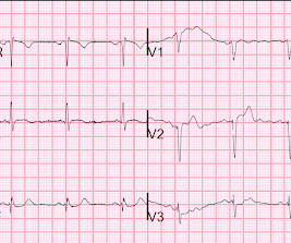
A middle aged male presented with chestpain. There may be ischemia present, but it is not evident on the ECG. In LVH, T-wave inversions are usually much more assymetric , like these (Figure 2): Acute Chestpain, but baseline ECG. Here is his ECG ( Figure 1 ): What do you think? All troponins were negative.
A 70-year-old man calls 911 after experiencing sudden, severe chestpain. Computer read: "Non-specific ST abnormality, consider anterior subendocardial ischemia" There are very poor R-waves in V1-V4 suggesting old anterior MI. Firstly, subendocardial ischemia does not localize on 12-Lead ECG. Circulation.
Submitted and written by Megan Lieb, DO with edits by Bracey, Smith, Meyers, and Grauer A 50-ish year old man with ICD presented to the emergency department with substernal chestpain for 3 hours prior to arrival. At this time he reported ongoing chestpain and was given aspirin and nitroglycerin. J Am Heart Assoc.
Of course this depends on many factors: 1) duration of occlusion, 2) whether full or near occlusion with zero flow or some flow -- the flow in the artery is the critical factor, measured by "TIMI" flow, 3) presence of collateral circulation and others. Upon questioning patient, he denies having any chestpain or chest tightness of any sort.
Circulation: Cardiovascular Imaging, Volume 16, Issue 11 , Page e015800, November 1, 2023. of patients had evidence of ischemia on a prior functional test. of patients had evidence of ischemia on a prior functional test. were referred to CCTA and 22.5% were referred to CCTA and 22.5% In the follow-up ranging from 1 to 3.5
This is where coronary circulation comes into play. Coronary circulation refers to the movement of blood through the network of coronary arteries and veins that supply the heart muscle (myocardium) itself. Step-by-Step Breakdown of Coronary Circulation 1.
Because the patient had no chestpain or shortness of breath, they were initially diagnosed as gastroenteritis. But because the patient had no chestpain or shortness of breath, it was not deemed to be from ACS. But because the patient had no chestpain or shortness of breath, it was not deemed to be from ACS.
Angiography usually reveals an absence of collateral circulation to the infarct zone. But in the other half of this 30% ( ie, in ~15% of all patients with MI ) — although these patients found on follow-up ECG to have had infarction did not have chestpain — they did have "something else" thought to be associated with their MI.
Written by Willy Frick A man in his 50s with a history of hypertension, dyslipidemia, type 2 diabetes mellitus, and prior inferior OMI status post DES to his proximal RCA 3 years prior presented to the emergency department at around 3 AM complaining of chestpain onset around 9 PM the evening prior. The following ECG was obtained.
Written by Jesse McLaren A 70 year old with prior MIs and stents to LAD and RCA presented to the emergency department with 2 weeks of increasing exertional chestpain radiating to the left arm, associated with nausea. Circulation 2014 2. But no ECG met STEMI criteria so the patient was referred to cardiology as Non-STEMI.
Jesse McLaren (@ECGcases), of Emergency Medicine Cases Reviewed by Pendell Meyers and Steve Smith An 85yo with a history of hypertension developed chestpain and collapsed, and had bystander CPR. On arrival, GCS was 13 and the patient complained of ongoing chestpain. Vitals were HR 58 BP 167/70 R20 sat 96%.
This male in his 40's had been having intermittent chestpain for one week. He awoke from sleep with crushing central chestpain and called ems. EMS recorded a 12-lead, then gave 2 sublingual nitros with complete relief of pain. Ischemia may be so brief that Wellens' waves do not evolve 3. Lessons: 1.
It was edited by Smith CASE : A 52-year-old male with a past medical history of hypertension and COPD summoned EMS with complaints of chestpain, weakness and nausea. The ECG cannot diagnose the etiology of ischemia; it only the presence of ischemia, from whatever etiology.
A middle aged male with no h/o CAD presented with one week of crescendo exertional angina, and had chestpain at the time of the first ECG: Here is the patient's previous ECG: Here is the patient's presenting ED ECG: There is isolated ST depression in precordial leads, deeper in V2 - V4 than in V5 or V6. There is no ST elevation.
This may result in ischemia (lack of oxygen to the heart muscle), causing parts of the heart to weaken and enlarge. Regular physical activity can strengthen the heart and improve circulation. Cardiomyopathy Cardiomyopathy is a condition that affects the heart muscle, causing it to become enlarged, thick, or rigid.
He denied chestpain or shortness of breath. In the clinical context of weakness and fever, without chestpain or shortness of breath, the likelihood of Brugada pattern is obviously much higher. Circulation, 117, 1890–1893. [3]: See below for PM Cardio digitized version of this. PM Cardio digitized version.
Circulation, Volume 150, Issue Suppl_1 , Page A4119267-A4119267, November 12, 2024. Advanced cardiac imaging especially in atypical presentations, can aid in early diagnosis.Case:A 59 year-old man with history of biopsy-proven pulmonary sarcoidosis presented with non exertional chestpain for 2 months.
But, in a patient who presents to the ED for new chestpain — seeing these subtle findings that are localized to leads V2- thru -V4 should at the least make you consider acute posterior OMI ( O cclusion-based MI ) — until you prove otherwise. To EMPHASIZE: These are subtle findings. What do YOU think?
The patient in today’s case is a previously healthy 40-something male who contacted EMS due to acute onset crushing chestpain. The pain was 10/10 in intensity radiating bilaterally to the shoulders and also to the left arm and neck. There is no definite evidence of acute ischemia. (ie, The below ECG was recorded.
This was a male in his 50's with a history of hypertension and possible diabetes mellitus who presented to the emergency department with a history of squeezing chestpain, lasting 5 minutes at a time, with several episodes over the past couple of months. Plan was for admission for chestpain workup. Gottlieb SO, et al.
This 42 yo diabetic male presented with cough and foot pain. In spite of aggressive questioning, he denied chestpain, but he did tell one triage nurse that he had had some chest burning, and so he underwent an ECG: There are deep Q-waves and QS-waves in precordial leads V2-V3, with a bit of R-wave left in V4.
However ,we have some effective clinical and pathological markers too, for effective re-vascularisation They are clinical well being and good functional capacity , relief from chest-pain, reduction of plaque volume, plaque stabilisation, maintenance of collaterals , microvascular patency , reduction of recurrent events.
Although the patient reported experiencing mild pressure-like chestpain, there was suspicion among clinicians that this might be indicative of an older change. The patient rapidly regained consciousness, reporting no residual pain. There is some ST-segment elevation in DII, DIII, aVF, V4-6.
He had no previous history of CAD, and presented with very typical waxing and waning chestpain, much worse with exertion but also present at rest and on presentation, though his pain was minimal at the time of the ECG. I saw this 59 year old male 3 weeks ago. Blood pressure was 150/80. The ECG normalized overnight.
A 70-something female with no previous cardiac history presented with acute chestpain. She awoke from sleep last night around 4:45 AM (3 hours prior to arrival) with pain that originated in her mid back. She stated the pain was achy/crampy. Over the course of the next hour, this pain turned into a pressure in her chest.
This fantastic case and post was written by Jesse McLaren (@ECGcases), edited by Smith Case You’re shown an ECG from a patient in the waiting room with chestpain. It was a 60yo with a history of stents to the circumflex and right coronary arteries, who presented with 9 hours of fluctuating central chestpain.
About this time, the 4th troponin, drawn at 8 hours after onset of pain, peaked at 20.956 ng/mL. Now you have ECG and troponin evidence of ischemia, AND ventricular dysrhythmia, which means this is NOT a stable ACS. It they are static, then they are not due to ischemia. Circulation. Again, cath lab was not activated.
Chest trauma was suspected on initial exam. The ECG shows sinus tachycardia with RBBB and LAFB, without clear additional superimposed signs of ischemia. Gunshot wound to the chest with ST Elevation Would your radiologist make this diagnosis, or should you record an ECG in trauma? Circulation: Cardiovascular Imaging.
Post by Smith and Meyers Sam Ghali ( [link] ) just asked me (Smith): "Steve, do left main coronary artery *occlusions* (actual ones with transmural ischemia) have ST Depression or ST Elevation in aVR?" That said, complete LM occlusion would be expected to have subepicardial ischemia (STE) in these myocardial territories: STE vector 1.
Case 3 : Male in 30's with chestpain, cough, and fever. Is there likely to be fixed coronary stenosis that led to demand ischemia during pneumonia? --Was Was the ST elevation due to transient demand ischemia, or is it false positive? What do you think? He has clinical pneumonia. Called 911. Does he need a stress test? --Is
This patient, who is a mid 60s female with a history of hypertension, hyperlipidemia and GERD, called 911 because of chestpain. A mid 60s woman with history of hypertension, hyperlipidemia, and GERD called 911 for chestpain. It is also NOT the clinical scenario of takotsubo (a week of intermittent chestpain).
There is no ischemia, certainly no concern at all for OMI. P.S.: Note that in today's case — the brief description of the chestpain history in this teenager does not sound convincing for an acute cardiac event. I see maybe one of these ECGs each month in my practice.
60-something with h/o MI and stents presented with chestpain radiating to the back and nausea/vomiting. For coronary anatomy, see here: [link] This is the post intervention ECG: All ST Elevation is gone (more proof that it was all a result of ischemia) Formal Echo: Normal estimated left ventricular ejection fraction - 55%.
There was no chestpain. Extensive conduction system abnormalities can have various causes (ischemia, genetic, infectious, amyloid, etc). The physiologic reason for this — is thought to be the result of momentarily increased circulation from mechanical contraction arising from the "sandwiched in" QRS complex.
Case A 39-year-old male without prior medical history presents with chestpain that started 2 hours prior to presentation. He says that the pain intensity was 10/10 at home but now about 4/10. Despite the clinical stability and decreasing pain, this patient needs an immediate angiogram. Circulation 2002; 105(4): 539-42.
Acute chestpain and a bizarre ECG Bizarre (Hyperacute??) Incredibly , this case was just published in Circulation on January 22, 2018 (thanks to Brooks Walsh for finding this!) link] Circulation. I thought the overall picture in these 6 chest leads did not look like acute OMI. What do you think? 2012;45(1):15-17.
A middle-aged woman had intermittent angina for 48 hours, then onset of constant, crushing chestpain for 1.5 More likely, the patient had crescendo angina, with REVERSIBLE ischemia for 48 hours that only became potentially irreversible (STEMI) at that point in time. Circulation 1993; 88:896-904. Methods: Oliva et al. (94)
It was from a patient with chestpain: Note the obvious Brugada pattern. Circulation, 117, 1890–1893. [3]: The elevated troponin was attributed to either type 2 MI or to non-MI acute myocardial injury. There is no further workup at this time. Smith: Here is a case that was just texted to me today from a former resident.
We organize all of the trending information in your field so you don't have to. Join thousands of users and stay up to date on the latest articles your peers are reading.
You know about us, now we want to get to know you!
Let's personalize your content
Let's get even more personalized
We recognize your account from another site in our network, please click 'Send Email' below to continue with verifying your account and setting a password.


Let's personalize your content