This site uses cookies to improve your experience. To help us insure we adhere to various privacy regulations, please select your country/region of residence. If you do not select a country, we will assume you are from the United States. Select your Cookie Settings or view our Privacy Policy and Terms of Use.
Cookie Settings
Cookies and similar technologies are used on this website for proper function of the website, for tracking performance analytics and for marketing purposes. We and some of our third-party providers may use cookie data for various purposes. Please review the cookie settings below and choose your preference.
Used for the proper function of the website
Used for monitoring website traffic and interactions
Cookie Settings
Cookies and similar technologies are used on this website for proper function of the website, for tracking performance analytics and for marketing purposes. We and some of our third-party providers may use cookie data for various purposes. Please review the cookie settings below and choose your preference.
Strictly Necessary: Used for the proper function of the website
Performance/Analytics: Used for monitoring website traffic and interactions
Coronary artery spasm (CAS), or Prinzmetal angina, is a recognised cause of myocardial ischaemia in non-obstructed coronary arteries which typically presents with anginal chestpain. The patient presented with recurrent palpitations and pre-syncope, with no chestpain.
In this ECG Cases blog, Jesse McLaren and Rajiv Thavanathan explore how ECG and POCUS complement each other for patients presenting to the emergency department with shortness of breath or chestpain. The post ECG Cases 49 – ECG and POCUS for Dyspnea and ChestPain appeared first on Emergency Medicine Cases.
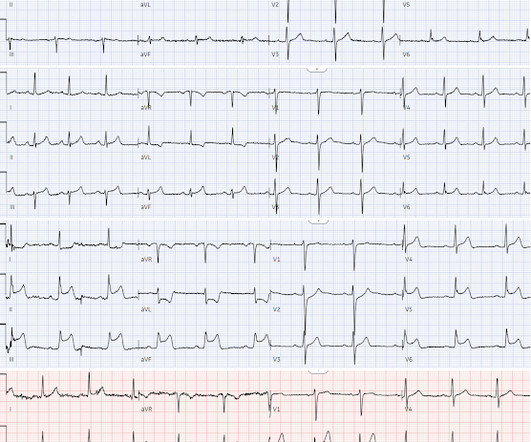
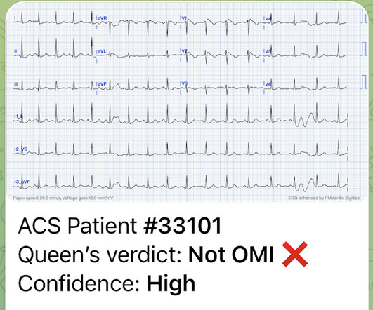
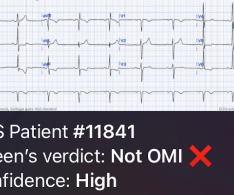
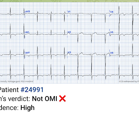
2 middle aged males presented with chestpain. Which had the more severe chestpain at the time of the ECG? Patient 2 at the bottom with a very subtle OMI complained of 10/10 chestpain at the time the ECG was recorded. 414 patients were included in the analysis.
Written by Jesse McLaren A 45-year-old presented with 24 hours of intermittent chestpain. On it’s own this is nonspecific, but in the right context this could be diagonal occlusion (if active chestpain) or infero-posterior reperfusion (if resolved chestpain). #2 Can you guess the sequence?
Written by Pendell Meyers Two patients with acute chestpain. Patient 1: Patient 2: Patient 1: A man in his 40s with minimal medical history presented with acute chestpain radiating to his R shoulder. Two patients with chestpain. Do either, neither, or both have OMI and need reperfusion?
I assumed it was a patient with acute chestpain. It was a man in his 30s with chestpain. This was sent to me from Sam Ghali ( @EM_Resus ) with no other information. What do you think, Steve? Real or just fake?" What do YOU think? It has some inferior ST elevation with some reciprocal ST depression and inverted T in aVL.
Written by Jesse McLaren, with a very few edits by Smith A 60-year-old presented with chestpain. Inferior hyperacute T waves, which have been added to the 2022 ACC consensus on chestpain as a “STEMI equivalent”[3] 3. But are there any other signs of Occlusion MI? Conduction disorders in the setting of acute STEMI.
Of course he said: "Yes, it was a 60 year old diabetic with Chestpain." K en G rauer gives a thorough explanation here: A 60 year old with chestpain == MY Comment , by K EN G RAUER, MD ( 9/15 /2023 ): == The 1st time that I saw APTA ( A rterial P ulse T ap A rtifact ) — I did not know what it was. That is not a STEMI.
A 50-something male had onset of chestpain 1 hour prior to ED arrival. Endorses some associated SOB, but denies back pain, fever, cough, chills, leg swelling, or other new symptoms. Always get serial ECGs in a patient with acute chestpain. It is constant, 9/10, left-sided CP that radiates into left arm and jaw.
A young woman presented with acute chestpain. This case came from a friend whose sister was the patient. She knew I was interested in ECGs, so she took a photo of this one. This was her presenting ECG: What do you think? This is clearly Brugada phenotype. There is downsloping ST Elevation in V1 and V2.
A man in his mid-50s presented with chestpain lasting 30 minutes. The initial electrocardiogram showed type A preexcitation syndrome, with obvious ST-segment depression in leads V3 through V5 and positive delta wave. What would you do next?
The patient was a middle-aged female who had acute chestpain of approximately 6 hours duration. The pain was still active at the time of evaluation. See some relevant cases below: Chestpain with anterior ST depression: look what happens if you use posterior leads.
ACC's ChestPain Center Accreditation is designed to help facilities to establish consistent, high-quality processes for the most efficient and effective acute coronary syndrome (ACS) care.
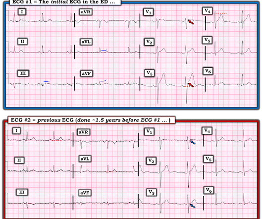
Written by Jesse McLaren A 45 year old presented with two weeks of recurring non-exertional chestpain, now constant for an hour. Because of the ECG changes in a patient with chestpain, and with inferolateral hypokinesis on POCUS, the cath lab was activated. Below is old and then new ECG (old on top; new below).
Written by Willy Frick A man in his early 40s with BMI 36, hypertension, and a 30 pack-year smoking history presented with three days of chestpain. He described it as a mild intensity, nagging pain on the right side of his chest with nausea and dyspnea. It started while he was at rest after finishing a workout.
Written by Willy Frick A 67 year old man with a history of hypertension presented with three days of chestpain radiating to his back. Due to the chestpain radiating into the patient's back, the ER physician ordered CTA chest to rule out aortic dissection. He had associated nausea, vomiting, and dyspnea.
Written by Jesse McLaren A healthy 75 year old developed 7/10 chestpain associated with diaphoresis and nausea, which began on exertion but persisted. Below is the first ECG recorded by paramedics after 2 hours of chestpain, interpreted by the machine as “possible inferior ischemia”. What do you think?
By Magnus Nossen This ECG is from a young man with no risk factors for CAD, he presented with chestpain. The patient is a young adult male with chestpain. The chestpain was described as pressure like and radiation to both arms and the jaw. How would you assess this ECG? What is your next step?
Sent by Magnus Nossen MD, written by Pendell Meyers A man in his 50s, previously healthy, developed acute chestpain. The primary care physician there evaluated this patient and deemed the chestpain to be due to gastrointestinal causes. The ECG was also interpreted as normal by the primary care physician.
Future heart attacks could be better prevented in people visiting their GP with unexplained chestpain, after Keele researchers developed the clearest picture yet of the factors that put them at higher risk. The research is published in the European Journal of Preventive Cardiology.
Written by Pendell Meyers A man in his 60s presented with acute chestpain and normal vital signs. Here is his triage ECG: What do you think? The ECG shows massively hyperacute T waves of LAD OMI, plus WPW. V3-V5 also have the depressed HATW takeoff which qualifies them as the rare de Winter subtype of HATWs.
Sent by anonymous, written by Pendell Meyers, reviewed by Smith and Grauer A man in his 40s presented to the ED with HTN, DM, and smoking history for evaluation of acute chestpain. He was eating lunch when he had sudden onset chest pressure, 9/10, radiating to his back, with sweating and numbness in both hands.
A 50 year old presented to the emergency department of a remote rural community (where the nearest cath lab is a plane ride away) with one hour of mild chestpain radiating to the back and jaw, and an ECG labeled ‘normal’ by the computer interpretation. What do you think, and how would you manage the patient?
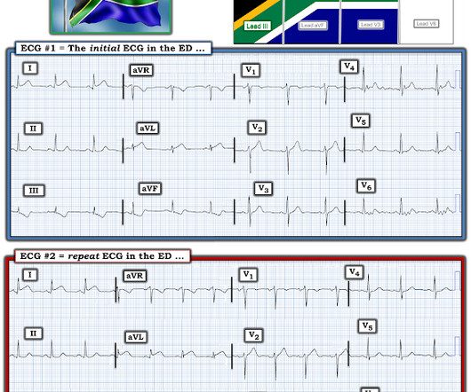
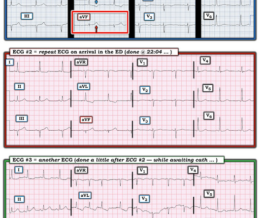
Written by Magnus Nossen with Edits by Grauer and Smith The ECGs in today’s case are from 3 different patients all presenting with new-onset CP ( ChestPain ). All ECGs were recorded by EMS, and transferred to a PCI capable center for evaluation. For 2 of the 3 patients — the cath lab was activated based on the ECG.
A 56 year old male with PMHx significant for hypertension had chestpain for several hours, then presented to the ED in the middle of the night. He reported chestpain that developed several hours prior to arrival and was 5/10 in intensity. The pain was located in the mid to left chest and developed after riding his bike.
(MedPage Today) -- For lower-risk patients with acute myocardial injury already ruled out for their chestpain, an increase in referrals for noninvasive cardiac testing (NICT) was not associated with improved outcomes, a retrospective cohort study.
This was sent by Sam Ghali @EM_RESUS A 44 year old man presented with chestpain The tech came running with the ECG as the computer called "STEMI!" What do you think? Sam sent this to me and asked: "What do you think, Steve?" My answer: --Tough one! --But
Written by Jesse McLaren A 65 year old with a history of atrial flutter, CABG and end-stage renal disease on dialysis presented with 3 days of fluctuating chestpain, which was ongoing at triage. So a patient with high pretest probability (prior CABG with new chestpain), had new ECG changes showing posterior OMI.
A 41-year-old male who presents to the emergency department with chestpain. Patient reports approximately 2 hours prior to arrival he developed a sharp chestpain that radiates into his left arm and left lower leg. Describes the radiating pain as numbness/tingling. No shortness of breath. No recent travel.
A 50-something male with hypertension and 20- to 40-year smoking history presented with 1 week of stuttering chestpain that is worse with exertion, which takes many minutes to resolve after resting and never occurs at rest. At times the pain does go to his left neck. It is a ssociated with mild dyspnea on exertion.
The patient presented to an outside hospital An 80yo female per triage “patient presents with chestpain, also hurts to breathe” PMH: CAD, s/p stent placement, CHF, atrial fibrillation, pacemaker (placed 1 month earlier), LBBB. HPI: Abrupt onset of substernal chestpain associated with nausea/vomiting 30 min PTA.
No ChestPain, but somnolent. The fact that this is syncope makes give it a far lower pretest probability than chestpain, but it was really more than syncope, as the patient actually underwent CPR and had hypotension on arrival of EMS. Here is the ED ECG (a photo of the paper printout) What do you think?
I was working at triage when the medics brought this patient who is 65 yo and has had chestpain for 12 hours. They recorded a prehospital ECG at 2112 and said that it was “normal”. It had already been crumpled up and put in the waste basket. So I uncrumpled it: What do you think?
Written by Willy Frick A man in his 50s with history of hypertension, hyperlipidemia, and a 30 pack-year smoking history presented to the ER with 1 hour of acute onset, severe chestpain and diaphoresis. His ECG is shown: What do you think? What do you think?
Written by Jesse McLaren Four patients presented with chestpain. All initial ECGs were labeled ‘normal’ or ‘otherwise normal’ by the computer interpretation, and below are the ECGs with the final cardiology interpretation.
A 54-year-old male athlete was evaluated at this hospital because of exertional dyspnea and chestpain. Physical examination revealed jugular venous distention and Kussmauls sign. A diagnosis was made.
(MedPage Today) -- PARIS -- Whether a person had chestpains resolved by angioplasty hinged on the nature, not the severity, of their presenting symptoms, an ORBITA-2 analysis showed. Investigators found two groups more likely to benefit from.
Written by Jesse McLaren, comments by Smith A 55 year old with a history of NSTEMI presented with two hours of exertional chestpain, with normal vitals. See these posts: ChestPain, ST Elevation, and an Elevated Troponin: Should we Activate the Cath Lab? What do you think?
Written by Jesse McLaren Two patients in their 70s presented to the ED with chestpain and RBBB. Patient 1 : a 75 year old called paramedics with one day of left shoulder pain which migrated to the central chest, which was worse with deep breaths. Do either, both, or neither have occlusion MI? Vitals were normal.
Written by Jesse McLaren Three patients presented with acute chestpain and ECGs that were labeled by the computer as completely normal, and which was confirmed by the final cardiology interpretation (which is blinded to patient outcome) also as completely normal. What do you think?
I went to the patient's chart: Elderly woman with stuttering chestpain and SOB, and dizziness. I was reading ECGs on the system and saw this one, and instantly knew the probable ECG diagnosis: What do you think? What do you think now? This is a very typical ECG for Hypertrophic Cardiomyopathy.
Written by Jesse McLaren A 50 year old presented to triage with one hour of chestpain, and the following ECG labeled normal by the computer (GE Marquette SL) algorithm. What do you think? Theres normal sinus rhythm, first degree AV block, early R wave, normal voltages. Here is her ECG: What do you think?
We organize all of the trending information in your field so you don't have to. Join thousands of users and stay up to date on the latest articles your peers are reading.
You know about us, now we want to get to know you!
Let's personalize your content
Let's get even more personalized
We recognize your account from another site in our network, please click 'Send Email' below to continue with verifying your account and setting a password.


Let's personalize your content