This site uses cookies to improve your experience. To help us insure we adhere to various privacy regulations, please select your country/region of residence. If you do not select a country, we will assume you are from the United States. Select your Cookie Settings or view our Privacy Policy and Terms of Use.
Cookie Settings
Cookies and similar technologies are used on this website for proper function of the website, for tracking performance analytics and for marketing purposes. We and some of our third-party providers may use cookie data for various purposes. Please review the cookie settings below and choose your preference.
Used for the proper function of the website
Used for monitoring website traffic and interactions
Cookie Settings
Cookies and similar technologies are used on this website for proper function of the website, for tracking performance analytics and for marketing purposes. We and some of our third-party providers may use cookie data for various purposes. Please review the cookie settings below and choose your preference.
Strictly Necessary: Used for the proper function of the website
Performance/Analytics: Used for monitoring website traffic and interactions
These may include the utilization of ultrasound guidance for arterial access, optimization of sheath and catheter selection based on patient anatomy, and the adoption of patent hemostasis devices to minimize complications and enhance overall procedural outcomes.
These may include the utilization of ultrasound guidance for arterial access, optimization of sheath and catheter selection based on patient anatomy, and the adoption of patent hemostasis devices to minimize complications and enhance overall procedural outcomes.
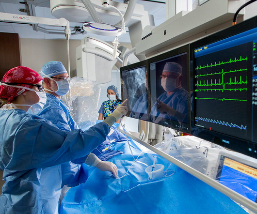
Discussing further, CatheterizationLaboratory, also called Cath Lab, is a medical examination room where angiogram, angioplasty, ablation, and implantation of pacemaker are carried out. Ultrasound, TEE, and IVUS play an influential role in dropping the usage of angiographic imaging.
Case continued A bedside cardiac ultrasound revealed grossly preserved left ventricular function, no appreciable wall motion abnormality, pericardial effusion, or obvious valvular abnormality. The terminal part of the T-wave is inverted in lead III, and reciprocally terminally upright in lead aVL.
We organize all of the trending information in your field so you don't have to. Join thousands of users and stay up to date on the latest articles your peers are reading.
You know about us, now we want to get to know you!
Let's personalize your content
Let's get even more personalized
We recognize your account from another site in our network, please click 'Send Email' below to continue with verifying your account and setting a password.


Let's personalize your content