This site uses cookies to improve your experience. To help us insure we adhere to various privacy regulations, please select your country/region of residence. If you do not select a country, we will assume you are from the United States. Select your Cookie Settings or view our Privacy Policy and Terms of Use.
Cookie Settings
Cookies and similar technologies are used on this website for proper function of the website, for tracking performance analytics and for marketing purposes. We and some of our third-party providers may use cookie data for various purposes. Please review the cookie settings below and choose your preference.
Used for the proper function of the website
Used for monitoring website traffic and interactions
Cookie Settings
Cookies and similar technologies are used on this website for proper function of the website, for tracking performance analytics and for marketing purposes. We and some of our third-party providers may use cookie data for various purposes. Please review the cookie settings below and choose your preference.
Strictly Necessary: Used for the proper function of the website
Performance/Analytics: Used for monitoring website traffic and interactions
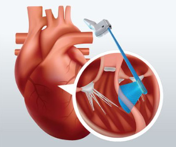
Traditionally, a cardiothoracic surgeon uses transesophageal echocardiography (TEE) before performing septal myectomy. The OPIE transducer is compatible with Fujifilm’s premium ultrasound system, the ARIETTA Precision. Currently, TEE cannot be leveraged during cardiac bypass surgery.
In this study, we compared the analgesic effects of intercostal nerve block (ICNB), ultrasound-guided paravertebral nerve block (PVB), and epidural block (EB) following single-port thoracoscopic lung surgery.
Goar received the recognition during a special ceremony in Paris, France, along with esteemed cardiothoracic surgeon, Professor Ottavio Alfieri of San Raffaele Hospital in Milan, Italy. He also played a key role in developing intravascular ultrasound, as well as the U.S.
Seeing two brothers have HLHS is extremely rare and even more rare is how differently the condition presented in each child, says Childrens Hospital Colorado cardiothoracic surgeon Max Mitchell, MD, who performed the procedures on Zane and advised on Zekes care.
Ultrasound-guided percutaneous axillary vein cannulation can reduce cannulation failure and mechanical complications, is as safe and effective as internal jugular vein cannulation, and is superior to subclavia.
Hemodynamic instability in trauma is usually due to bleeding, but if ultrasound shows poor contractility, then this may be due to cardiac contusion. In the ED, ultrasound showed hemopericardium with tamponade. Outcome Three weeks later, shortly after having been physically active (bouncing on a trampoline), she was found unresponsive.
It was notable for a normal cardiac ultrasound with no pericardial fluid, normal LV and RV function (though the quality was not sufficient to evaluate for wall motion abnormalities) and normal IVC dynamics. Bedside ultrasound is another very important piece. Ultrasound can be very helpful to distinguish causes of hypotension.
The lung ultrasound score (LUS) was assessed following anesthesia and resuscitation (T5). The PaO2:FiO2 ratio was measured using blood gas analysis 30 min after initiating one-lung breathing (T2) and immediately when OLV ended (T3). The occurrence of atelectasis was documented immediately after the surgery.
We organize all of the trending information in your field so you don't have to. Join thousands of users and stay up to date on the latest articles your peers are reading.
You know about us, now we want to get to know you!
Let's personalize your content
Let's get even more personalized
We recognize your account from another site in our network, please click 'Send Email' below to continue with verifying your account and setting a password.


Let's personalize your content