This site uses cookies to improve your experience. To help us insure we adhere to various privacy regulations, please select your country/region of residence. If you do not select a country, we will assume you are from the United States. Select your Cookie Settings or view our Privacy Policy and Terms of Use.
Cookie Settings
Cookies and similar technologies are used on this website for proper function of the website, for tracking performance analytics and for marketing purposes. We and some of our third-party providers may use cookie data for various purposes. Please review the cookie settings below and choose your preference.
Used for the proper function of the website
Used for monitoring website traffic and interactions
Cookie Settings
Cookies and similar technologies are used on this website for proper function of the website, for tracking performance analytics and for marketing purposes. We and some of our third-party providers may use cookie data for various purposes. Please review the cookie settings below and choose your preference.
Strictly Necessary: Used for the proper function of the website
Performance/Analytics: Used for monitoring website traffic and interactions
Specific genetic variants, such as those affecting cholesterol metabolism, can increase the likelihood of plaque buildup in the arteries. Cardiomyopathies: These diseases affect the heart muscle, impairing its ability to pump blood effectively.
Because it’s the month that houses Valentine’s Day, it is obviously the most appropriate month to share information about takotsubo cardiomyopathy. Takotsubo cardiomyopathy is commonly known by a few other names as well – stress cardiomyopathy, apical ballooning syndrome, broken heart syndrome, and stress-induced cardiomyopathy.
Angiogram No obstructive epicardial coronary artery disease Cannot exclude non-ACS causes of troponin elevation including coronary vasospasm, stress cardiomyopathy, microvascular disease, etc. We know that most type 1 acute MI due to plaque rupture and thrombosis occurs in lesions that are less than 50% (see Libby reference).
Coronary Artery Disease (CAD) CAD, which involves the narrowing or blockage of coronary arteries due to plaque buildup, can reduce blood flow to the heart. CardiomyopathyCardiomyopathy is a condition that affects the heart muscle, causing it to become enlarged, thick, or rigid.
Non-obstructive coronary disease at the time cardiac cath is done does not necessarily imply there was no plaque rupture with thrombus. These plaques will often not be recognized as "culprits" — because no fissuring or ulceration is seen. Longterm prognosis of patients with MINOCA clearly depends on the underlying etiology.
The commonest causes of MINOCA include: atherosclerotic causes such as plaque rupture or erosion with spontaneous thrombolysis, and non-atherosclerotic causes such as coronary vasospasm (sometimes called variant angina or Prinzmetal's angina), coronary embolism or thrombosis, possibly microvascular dysfunction. It is not rare.
Given her lack of risk factors for coronary disease, and the fact that she was a 53 year old woman with compatible history and echo findings, stress cardiomyopathy rose to the top of my differential. Of course, stress cardiomyopathy is a diagnosis of exclusion. But not all OMI is atherosclerotic in nature.
Learning Points A fantastic triage tool for syncope is the ECG, which can screen for any arrhythmic / structural contribution, such as Delta wave, QT prolongation, AV block, Hypertrophic Cardiomyopathy, Epsilon wave, or Brugada. The underlying etiology is either Type 1 or Type II ischemia, although sometimes there’s overlap of both.
Awards granted: a) Plaque for each awarded article. The corresponding recognition will entitle the honoree to a plaque. The Award will correspond to articles published in 2023. b) Registration for the next subsequent congress. c) Credit of R$ 2,000.00 for enrollment in courses at the Heart University. Except for postgraduate courses.
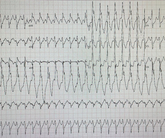
I interpreted the ECG as VT with two primary etiological possibilities: 1. Abrupt plaque ulceration of Type 1 ACS leading to VT. 2. Baseline fibrotic substrate from dilated cardiomyopathy leading to VT. From a clinical standpoint I reconciled the first possibility as most probable since the chest discomfort came first.
by making it clear to everyone that this is NOT an EKG that one sees with takotsubo cardiomyopathy. Hospital Course The patient was taken emergently to the cath lab which did not reveal any significant coronary artery disease, but she was noted to have reduced EF consistent with Takotsubo cardiomyopathy. It can only be seen by IVUS.
Example here: DIffuse ST Elevation with Apical Ballooning: is it Takotsubo Stress Cardiomyopathy? Mild Plaque no angiographically significant obstructive coronary artery disease. Stress induced cardiomyopathy (Takotsubo like LV dysfunction) possible. Therefore, the cath lab was activated. The initial hs troponin was = 41 ng/L.
If our valves are leaky then again, a smaller volume of blood is effectively ejected because some leaks back Cardiomyopathies – if the heart muscle itself is defective in some way then that makes it weaker. This includes conditions like familial hypertrophic cardiomyopathy, familial dilated cardiomyopathy etc.
For instance, younger (less than 30 or 40 years of age) individuals who suffer from sudden cardiac death during exercise are usually found to have an inherited or congenital cardiac condition, also known collectively as cardiomyopathies. Indeed, 44% of deaths among athletes in the U.S. ” Fear-inducing for heavy exercisers?
Patients with obstructive hypertrophic cardiomyopathy who underwent surgical myectomy reported improved quality of life. JACC: Asia) Lexaria Bioscience has announced that a CBD product beats a placebo in simulating acute pulmonary hypertension.
She had some very minor plaque but certainly nothing that could explain the heart attack and therefore she was discharged with a diagnosis of MINOCA i.e Then I think it is important that patient has an assessment of the function of the heart by means of an ultrasound to look for cardiomyopathies, Takotsubo etc.
Patients receiving semaglutide showed a greater change in Kansas City Cardiomyopathy Questionnaire (KCCQ) clinical summary scores at 52 weeks than placebo. These patients were identified to have non-flow-limiting vulnerable coronary plaques through intracoronary imaging.
The combination of prolonged QT and deep T wave inversion throughout the precordium is typical of Takotsubo syndrome, or Stress Cardiomyopathy – which can occur in the context of a physiologically distressed ICU patient, further compromising their hemodynamics. The coronary angiogram revealed no critical stenosis, or acute plaque ulceration.
No family history of sudden cardiac death, cardiomyopathy, premature CAD, or other cardiac issues. Pattern consistent with Takotsubo's cardiomyopathy." Only after her troponin peaked at 500,000 ng/L did she get her angiogram, which showed a 100% left main occlusion due to ruptured plaque. No similar symptoms in the past.
An ECG machine is able to detect other abnormalities of the heart as well, such as hypertrophic cardiomyopathy or overly thick heart muscles. Coronary artery disease Excessive cholesterol builds up plaque that blocks the arteries supplying blood to the heart. ECG tests can also show previous heart attacks.
plaque disruption), the T waves still manifest markings of a previous state of suboptimal coronary flow that resolved: Type II supply-demand mismatch in the setting of extreme bradycardia. LBBB is typically the result of preexisting hypertrophy, ischemic heart disease, or cardiomyopathy. 5] Isnard, R. & Pousset, F. 6] Tabrizi, F.,
So cardiomyopathies, valve problems, myocarditis and previous heart attacks all cause a problem with the pumping function of the heart. If the pump is in any way defective then not as much blood comes out of the heart and the body and all our vital organs will get less blood than they should and this could lead to harm.
Troponins may be negative with very rapid reperfusion, or measured too late, or chronically elevated due to cardiomyopathy or renal failure. Angiograms may be negative due to spasm or thrombus lysis or small vessel disease, or it may be a type 2 MI. SCAD occurs in patients with few or non-traditional cardiovascular risk factors.
Repeat ECG was obtained immediately, just 24 minutes after the prior ECG: Given the context, my top differential diagnosis would be stress cardiomyopathy AKA takotsubo. In my opinion, the more likely explanation is that the ST-T changes are primarily driven by stress cardiomyopathy. Here is a case report and review of the literature.
Whenever I see PVCs with the morphology and axis seen in todays case I always look for signs of AC ( Arrhythmogenic Cardiomyopathy ). Arrhythmogenic cardiomyopathy often manifests with PVCs from the RV. The ECG in Figure-1 however, shows no signs of arrhythmogenic cardiomyopathy. There were no plaques or stenoses.
We organize all of the trending information in your field so you don't have to. Join thousands of users and stay up to date on the latest articles your peers are reading.
You know about us, now we want to get to know you!
Let's personalize your content
Let's get even more personalized
We recognize your account from another site in our network, please click 'Send Email' below to continue with verifying your account and setting a password.


Let's personalize your content