This site uses cookies to improve your experience. To help us insure we adhere to various privacy regulations, please select your country/region of residence. If you do not select a country, we will assume you are from the United States. Select your Cookie Settings or view our Privacy Policy and Terms of Use.
Cookie Settings
Cookies and similar technologies are used on this website for proper function of the website, for tracking performance analytics and for marketing purposes. We and some of our third-party providers may use cookie data for various purposes. Please review the cookie settings below and choose your preference.
Used for the proper function of the website
Used for monitoring website traffic and interactions
Cookie Settings
Cookies and similar technologies are used on this website for proper function of the website, for tracking performance analytics and for marketing purposes. We and some of our third-party providers may use cookie data for various purposes. Please review the cookie settings below and choose your preference.
Strictly Necessary: Used for the proper function of the website
Performance/Analytics: Used for monitoring website traffic and interactions
A 69-year-old man with a history of MI and cardiomyopathy presented with 2 days of dyspnea. Regular tachycardia (124 beats per min), diaphoresis, and rales were present. A diagnosis was made.
In this review, we comprehensively review the regulation mechanism of SFⅡ helicases in CVDs including atherosclerosis, myocardialinfarction, cardiomyopathies, and heart failure, which will contribute to the investigation of ideal therapeutic targets for CVDs.
She reports a known history of Hypertrophic Cardiomyopathy (HCM) with left ventricular outflow tract obstruction and is on daily beta blocker therapy. Diagnosis and management of hypertrophic cardiomyopathy: Expert analysis. Chapter 10: Hypertrophic Cardiomyopathy (pg. References Naidu, S. American College of Cardiology.
Hypertrophic cardiomyopathy is a genetic disorder with a guarded prognosis which occurs in about 1:500 individuals. The most common symptom of hypertrophic cardiomyopathy is dyspnoea which occurs in 90% of cases and is due to elevated left ventricular diastolic pressures as a consequence of the diastolic dysfunction.
Angiogram No obstructive epicardial coronary artery disease Cannot exclude non-ACS causes of troponin elevation including coronary vasospasm, stress cardiomyopathy, microvascular disease, etc. Medical Therapy for Secondary Prevention and Long-Term Outcome in Patients With MyocardialInfarction With Nonobstructive Coronary Artery Disease.
The intergroup comparison showed the therapeutic effect of the combination was equivalent in HF caused by myocardialInfarction (MI), dilated cardiomyopathy (DCM) or Valvular Heart Disease (VHD).ConclusionVericiguat and improved patients’ quality of life scores.
This is without any intervening infarction. He has "ischemic cardiomyopathy" and "congestive heart failure." In the present STEMI/NSTEMI paradigm, these 2 greatly different (heterogeneous) infarcts are lumped together as one kind of infarct! And this is after undergoing very rapid intervention.
This comprehensive approach allows for precise differentiation between various cardiomyopathies, myocarditis, and myocardialinfarction. Hundley describes that CMR has become a “one-stop shop” for evaluating cardiovascular morphology, perfusion, function, and tissue characterization in a single examination.
Protein aggregates are emerging therapeutic targets in rare monogenic causes of cardiomyopathy and amyloid heart disease, but their role in more prevalent heart-failure syndromes remains mechanistically unexamined. 25-Hydroxycholesterol attenuated CRYAB serine 59 phosphorylation and rescued postmyocardial infarction adverse remodeling.
by making it clear to everyone that this is NOT an EKG that one sees with takotsubo cardiomyopathy. Hospital Course The patient was taken emergently to the cath lab which did not reveal any significant coronary artery disease, but she was noted to have reduced EF consistent with Takotsubo cardiomyopathy. It can only be seen by IVUS.
This study aimed to assess the association between PVC burdens during submaximal exercise and major adverse cardiovascular events (MI/HF/LTVA: myocardialinfarction [MI], heart failure [HF], and life-threatening ventricular arrhythmia [LTVA]), and all-cause mortality.
See our publication: ST depression in lead aVL differentiates inferior ST-elevation myocardialinfarction from pericarditis There is STE in inferior leads, high lateral leads, and V4-V6. When there is ST depression (as in aVL, V2, V3), then top on the differential is OMI or myocarditis.
So this is indeed diagnostic of myocardialinfarction. Not immediately, at least, because this is NOT diagnostic of ACUTE (occlusion) myocardialinfarction (Acute OMI). So I made an ED diagnosis of Non-Occlusion MyocardialInfarction (NOMI), and his next day angiogram confirmed NOMI.
The term MINOCA stands for Myocardialinfarction with non-obstructive coronary arteries. Then I think it is important that patient has an assessment of the function of the heart by means of an ultrasound to look for cardiomyopathies, Takotsubo etc. I’ll try and explain this a bit better by using a case study.
This is diagnostic of myocardialinfarction. Troponins may be negative with very rapid reperfusion, or measured too late, or chronically elevated due to cardiomyopathy or renal failure. She felt more comfortable being admitted. Subsequent events: Later, before being taken to her room, the 2nd troponin returned at 1.01
Alternatively , it is someone who has an old myocardialinfarction and is now very sick with something else. He had a h/o ischemic cardiomyopathy and right MCA stroke. Below is my response. There is sinus tach. There is an incomplete RBBB and LAFB. There are QR-waves in aVL and V2, and a QS-wave in V3.
The important point for our purposes is that they do no represent myocardialinfarction. Hypertrophic Cardiomyopathy or Normal ("Variant")? Dr. Smith note: I wouldn't necessarily consider this ENTIRELY "benign." It may even be that patients with ECGs with this morphology have a higher long term risk.
A triphasic left ventricular filling pattern with an additional mid diastolic wave, called T wave by some authors and L wave by others, can occur in situations of left ventricular diastolic dysfunction, especially in hypertrophic cardiomyopathy. Another video on this channel describes triphasic mitral flow in more detail. Møller JE et al.
He reported a history of ischemic cardiomyopathy with coronary stent placement approximately 10 years prior, but could not recall the specific artery involved. Terminal QRS distortion is present in anterior myocardialinfarction but absent in early repolarization. Attached is the first ECG. 3] Smith, S. 4] Baranchuk, A, et al.
Next trop 85,528 ng/L (consistent with a massive myocardialinfarction), none further measured. LCX: minimal luminal irregularities RCA: minimal luminal irregularities Aspiration thrombectomy and PCI performed with resultant TIMI 3 flow in LAD and D1. 1824 post cath: Mot much different than the ECG above, but QOH: "OMI, Low confidence."
BackgroundStrain assessed by cardiac magnetic resonance (CMR) is a key prognostic indicator in myocardialinfarction. However, the strain characteristics and prognostic value in myocardialinfarction with nonobstructive coronary arteries (MINOCA) with different causes are unclear.
Arrhythmogenic Cardiomyopathy was better known as Arrhythmogenic Right Ventricular Dysplasia or ARVD and sometimes as Arrhythmogenic Right Ventricular Cardiomyopathy or ARVC earlier. A study published in JACC has compared the phenotypic expression and clinical outcomes in patients with arrhythmogenic cardiomyopathy [2].
Because it’s the month that houses Valentine’s Day, it is obviously the most appropriate month to share information about takotsubo cardiomyopathy. Takotsubo cardiomyopathy is commonly known by a few other names as well – stress cardiomyopathy, apical ballooning syndrome, broken heart syndrome, and stress-induced cardiomyopathy.
The noradrenergic phenotype had significantly more atherosclerotic complications (composite end point of type 1 myocardialinfarction and symptomatic peripheral artery disease; odds ratio, 3.58 [95% CI, 1.59–8.83];P=0.003), 0.57];P=0.002).
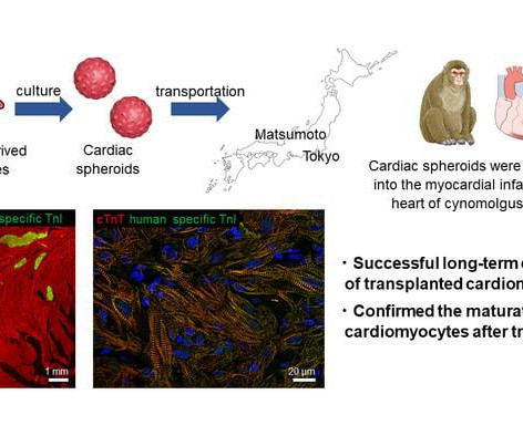
Myocardialinfarctions , commonly known as “heart attacks,” are on the rise, resulting in a significant number of deaths each year. We are already employing the same cardiac spheroids on patients with ischemic cardiomyopathy,” comments Asst.
Sudden cardiac death (SCD) risk stratification is based on clinically recognized risk factors (RF), such as reduced left ventricular (LV) ejection fraction (EF), heart failure (HF), prior myocardialinfarction (MI), and syncope. These RFs fail to capture the majority of SCDs.
BackgroundRecent studies have indicated high rates of future major adverse cardiovascular events in patients with Takotsubo cardiomyopathy (TC), but there is no well‐established tool for risk stratification. Journal of the American Heart Association, Ahead of Print.
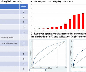
Background Takotsubo cardiomyopathy (TC) is an established differential diagnosis of myocardialinfarction with non-obstructive coronaries with significant interest but limited data on prognostication. We reviewed the characteristics and in-hospital outcomes and developed a novel risk score for TC.
Within the last six months, separate AI-ECG algorithms for detecting Low Ejection Fraction (Anumana), Hypertrophic cardiomyopathy (Viz.ai), and Occlusion Myocardialinfarction (Powerful Medical) have all been granted regulatory clearance (the latter under the EU MDR) and are in the early stages of deployment.
In this study, two independent cohorts were used to investigate for cardiovascular magnetic resonance (CMR) and genetic features of dilated cardiomyopathy (DCM) in individuals with coronary artery disease (CAD). Methods and results This study includes two cohorts.
Impella Left Sided Blood Pumps also are used when there is ongoing cardiogenic shock that occurs less than 48 hours after a severe heart attack (acute myocardialinfarction), open-heart surgery, or when the heart is not functioning well due to a condition called cardiomyopathy.
Background:Clinical and genetic predispositions are significant in predicting atrial fibrillation (AF); however, their role in patients with hypertrophic cardiomyopathy (HCM) remains unclear. Stroke, Volume 56, Issue Suppl_1 , Page ATMP101-ATMP101, February 1, 2025.
Scar burden on LGE-CMR imaging may be a risk marker for ventricular arrhythmia post-myocardialinfarction (MI). There is an unmet need for better risk stratification for sudden cardiac death in the era of primary prevention ICD therapy.
BackgroundHeart failure is a common complication after myocardialinfarction (MI) and is associated with increased mortality. Whether remote heart failure symptoms assessment after MI can improve risk stratification is unknown.
We aimed to investigate the safety of intravenous tissue plasminogen activator (IV tPA) and mechanical thrombectomy (MT) for acute ischemic stroke (AIS), and compare the functional outcome in cardiomyopathy patients with and without HF. This finding was regardless of the type of intervention received, including tPA (15.9% 22.3%, p= 0.321).
Sudden heart failure has been well described after the death of a loved one and is known as Takatubo Cardiomyopathy after the shape of a Japanese octopus fishing pot. That event might have been a heart rhythm issue or even a cardiac arrest. This is more commonly known as ‘Broken Heart Syndrome.
Background Consensus guidelines support the use of implanted cardioverter-defibrillators (ICD) for primary prevention of sudden cardiac death in patients with either non-ischaemic or ischaemic cardiomyopathy with left ventricular ejection fraction (LVEF) ≤35%. vs 19.1%, p<0.0001) and myocardialinfarction (56.1%
Clin Chem [Internet] 2020;Available from: [link] Smith mini-review: Troponin in Emergency Department COVID patients Cardiac Troponin (cTn) is a nonspecific marker of myocardial injury. In normal times, the most common use of cTni is in diagnosing, or ruling out, acute myocardialinfarction (AMI, a subcategory of acute myocardial injury.
There is ST elevation in V2 and V3 There are inverted T-waves in V2 and V3 There are prominent U-waves in V2 and V3 Many responders were worried about ischemia or hypertrophic cardiomyopathy. Here was Massimo's response: "I'm very sure of Early Repolarization (ERP) diagnosis in this case. See this post on Benign T-wave Inversion.
And, addressing myocardialinfarction, recent studies have shown that 1-4% of Athletes untimely die due to myocarditis caused by COVID-19. Covid-19 and Risk of Myocarditis in Athletes: Recent Studies There is growing evidence that the SARS-CoV-2 version of the Covid-19 virus can directly infiltrate myocardial cells.
Detailed Considerations LBBB and MyocardialInfarction In the emergent setting it’s important to assess LBBB through the lens of the Smith-modified Sgarbossa criteria, especially in a context that is clinically consistent with Acute Coronary Syndrome. Left bundle branch block-induced cardiomyopathy: Myth or reality?
HIV and Cardiovascular Disease: HIV infection itself is associated with an increased risk of cardiovascular disease and can double the risk of CVDs, including myocardialinfarction and stroke. Plus, abacavir, an NRTI, has been linked to an increased risk of myocardialinfarction.
We organize all of the trending information in your field so you don't have to. Join thousands of users and stay up to date on the latest articles your peers are reading.
You know about us, now we want to get to know you!
Let's personalize your content
Let's get even more personalized
We recognize your account from another site in our network, please click 'Send Email' below to continue with verifying your account and setting a password.


Let's personalize your content