This site uses cookies to improve your experience. To help us insure we adhere to various privacy regulations, please select your country/region of residence. If you do not select a country, we will assume you are from the United States. Select your Cookie Settings or view our Privacy Policy and Terms of Use.
Cookie Settings
Cookies and similar technologies are used on this website for proper function of the website, for tracking performance analytics and for marketing purposes. We and some of our third-party providers may use cookie data for various purposes. Please review the cookie settings below and choose your preference.
Used for the proper function of the website
Used for monitoring website traffic and interactions
Cookie Settings
Cookies and similar technologies are used on this website for proper function of the website, for tracking performance analytics and for marketing purposes. We and some of our third-party providers may use cookie data for various purposes. Please review the cookie settings below and choose your preference.
Strictly Necessary: Used for the proper function of the website
Performance/Analytics: Used for monitoring website traffic and interactions
She had a single chamber ICD/Pacemaker implanted several years prior due to ventricular tachycardia. Answer : The ECG above shows a regular wide complex tachycardia. Said differently, the ECG shows a rather slow ventricular tachycardia with a 2:1 VA conduction. Cardiac output (CO) was being maintained by the tachycardia.
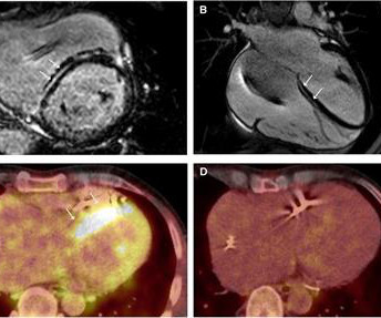
Cardiac sarcoidosis (CS), a rare condition characterized by non-caseating granulomas, can manifest with symptoms such as atrioventricular block and ventricular tachycardia (VT), as well as mimic inherited cardiomyopathies. A 58-year-old woman presented with sustained VT with a prior diagnosis of hypertrophic cardiomyopathy (HCM).
Studies have shown that mutations in the RYR2 gene, which encodes the RyR2 protein, are linked to several cardiac arrhythmias, including catecholaminergic polymorphic ventricular tachycardia (CPVT), long QT syndrome (LQTS), calcium release deficiency syndrome (CRDS), and atrial fibrillation (AF).
Atrial tachycardia (AT) originating from the left atrial appendage (LAA) is uncommon and the most difficult arrhythmia to eliminate. Therefore, we present the case of a 5-year-old girl with tachycardia-induced.
IntroductionFocal atrial tachycardia (FAT) is predominant in the pediatric population. A 12-lead electrocardiogram revealed a narrow QRS complex tachycardia with a rate of 157 beats per minute and a prolonged RP relationship. Echocardiography indicated a severely reduced ejection fraction of 22%.
Abstract Introduction Due to its unique features, pulsed field ablation (PFA) could potentially overcome some limitations of current radiofrequency (RF) ventricular tachycardia (VT) ablation. Methods Two patients with ischemic cardiomyopathy and previously failed RF VT ablations were treated with PFA.
Circulation: Arrhythmia and Electrophysiology, Ahead of Print. BACKGROUND:Epicardial approach in ventricular tachycardia (VT) ablation is still regarded as a second-step strategy, due to the risk of complications. The epicardial approach was considered useful if epicardial ablation was performed after epicardial mapping.
The advent of transseptal puncture has enabled the study of atrial fibrillation mechanisms, while epicardial access has enabled exploration of the epicardium in ventricular arrhythmias (ventricular tachycardia/ventricular fibrillation [VF]).1,2
Paco Dardon (@PacoDardon), and it’s a privilege to present it as a formal review due to the many pathophysiological, and electrophysiological, phenomenon at play. McLaren : ACLS attempts to simplify this process, suggesting cardioversion for unstable tachycardias, and anti-arrhythmics for stable wide complex tachycardias.
Chagas disease (ChD) was associated with increased rates of ventricular tachycardia and ventricular fibrillation in ICD patients only in the initial two periods, but there was no statistical difference in the last period. Progressive decline across periods in mortality rates among patients with implantable cardioverter-defibrillator (ICD).
Here is his 12-lead: There is a wide complex tachycardia with a rate of 257, with RBBB and LPFB (right axis deviation) morphology. Read about Fascicular VT here: Idiopathic Ventricular Tachycardias for the EM Physician Case Continued He was completely stable, so adenosine was administered. See Learning point 1 below. Arch Intern Med.
Fragmented QRS is a marker of myocardial scar and consequent arrhythmias in ischemic and nonischemic cardiomyopathy. Ebstein’s anomaly may be associated with right sided accessory pathway in ventricular pre-excitation, that is WPW syndrome, with or without atrioventricular re-entrant tachycardia.
Here was his ED ECG: There is sinus tachycardia (rate about 114) with nonspecific ST-T abnormalities. The absence of any wall motion abnormality makes ischemic cardiomyopathy very unlikely. An ECG was recorded: This shows a regular narrow complex tachycardia at a rate of about 160. BP:143/99, Pulse 109, Temp 37.2 °C
ABSTRACT Introduction Radiofrequency ablation is a cornerstone therapy for patients with ischemic cardiomyopathy (ICM) presenting with ventricular tachycardia (VT). In this context, ablation is typically performed endocardially as a first-line approach.
We organize all of the trending information in your field so you don't have to. Join thousands of users and stay up to date on the latest articles your peers are reading.
You know about us, now we want to get to know you!
Let's personalize your content
Let's get even more personalized
We recognize your account from another site in our network, please click 'Send Email' below to continue with verifying your account and setting a password.


Let's personalize your content