This site uses cookies to improve your experience. To help us insure we adhere to various privacy regulations, please select your country/region of residence. If you do not select a country, we will assume you are from the United States. Select your Cookie Settings or view our Privacy Policy and Terms of Use.
Cookie Settings
Cookies and similar technologies are used on this website for proper function of the website, for tracking performance analytics and for marketing purposes. We and some of our third-party providers may use cookie data for various purposes. Please review the cookie settings below and choose your preference.
Used for the proper function of the website
Used for monitoring website traffic and interactions
Cookie Settings
Cookies and similar technologies are used on this website for proper function of the website, for tracking performance analytics and for marketing purposes. We and some of our third-party providers may use cookie data for various purposes. Please review the cookie settings below and choose your preference.
Strictly Necessary: Used for the proper function of the website
Performance/Analytics: Used for monitoring website traffic and interactions
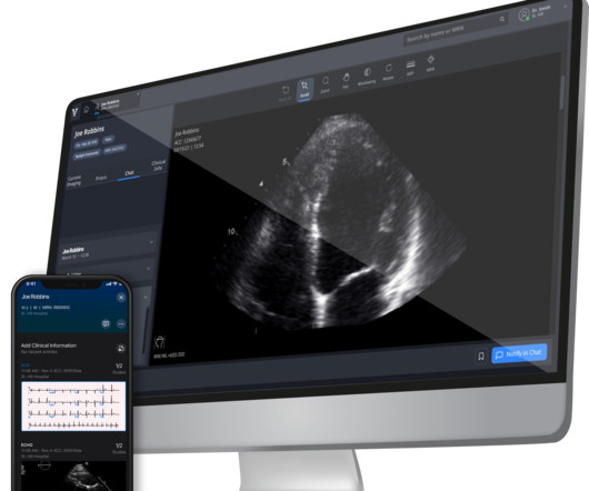
Researchers used an AI-enabled digital stethoscope that captures electrocardiogram ( ECG ) data and heart sounds to identify twice as many cases of peripartum cardiomyopathy as compared to regular care, according to a news release from the American Heart Association.
in clinical practice for patients with hypertrophic cardiomyopathy (HCM), a commonly inherited heart disease that often goes undetected. Three studies, presented at the American College of Cardiology ’s ( ACC ) Annual Scientific Session & Expo 2024 , have shown positive outcomes with real-world impact of Viz.ai sensitivity.
Because it’s the month that houses Valentine’s Day, it is obviously the most appropriate month to share information about takotsubo cardiomyopathy. Takotsubo cardiomyopathy is commonly known by a few other names as well – stress cardiomyopathy, apical ballooning syndrome, broken heart syndrome, and stress-induced cardiomyopathy.
Objectives In hypertrophic cardiomyopathy (HCM), specific ECG abnormalities are observed. In addition, the patients were divided into groups according to Kansas City Cardiomyopathy Questionnaire (KCCQ)-12. Therefore, ECG is a valuable screening tool. The model was trained in advance using PTB-XL, which is an open ECG dataset.
In patients undergoing non-physiologic pacemaker (PM) implantation (eg, RV pacing), cardiomyopathy (CM) may develop - particularly in patients with an elevated pacing burden. In contrast to the current standard of upgrading the pacemaker only once CM develops, a strategy to predict CM before it actually develops would be advantageous.
Whether you’re gearing up for your Family Medicine or Cardiology Board Exam, you’ll need to master the topic of Hypertrophic Cardiomyopathy (HCM). Explanation: Shown electrocardiogram suggests left ventricular hypertrophy. Hypertrophic cardiomyopathy is one of them. Hypertrophic cardiomyopathy is one of them.
Hypertrophic cardiomyopathy (HCM) has a prevalence of 1 in 500 people and highly increases the risk of sudden cardiac death (SCD). Diagnosis is typically based on the Echocardiogram, while cardiac MRI is used for detection of myocardial scarring through Late Gadolinium Enhancement (LGE), an important risk marker for SCD.
BackgroundRestrictive cardiomyopathy (RCM) is a rare cardiomyopathy often characterized by normal or reduced ventricular chamber volume and bi-atrial enlargement, caused mainly by mutations in the myonodal gene.
Thirty day electrocardiogram (ECG) monitoring in patients with hypertrophic cardiomyopathy (HCM) detects more arrhythmias than the standard 24 to 48 hours, according to late breaking science presented at EHRA 2023, a scientific congress of the European Society of Cardiology (ESC).
Diagnosis and assessment of disease progression in arrhythmogenic cardiomyopathy (ACM) are currently based on the 2010 Task Force Criteria (TFC) with the 12-lead electrocardiogram (ECG) being an important modality. The presence of inverted T-waves might be associated with lethal ventricular arrhythmias.
Cardiac sarcoidosis (CS) manifestations, often related to rhythm abnormalities and ventricular dysfunction, are non-specific and can mimic other cardiomyopathies.1
1 We hypothesized that the clinical data during the period of sinus maintenance would reflect the degree of atrial cardiomyopathy and could predict late arrhythmia recurrences during 12 months after CA of persistent AF.
CardiomyopathyCardiomyopathy is a condition that affects the heart muscle, causing it to become enlarged, thick, or rigid. Excessive Alcohol or Drug Use Long-term abuse of alcohol or certain drugs can weaken the heart muscle, resulting in cardiomyopathy and eventually cardiomegaly.
Artificial intelligence (AI)-enabled electrocardiography (ECG) scores can predict atrial fibrillation (AF) and grade atrial cardiomyopathy. Advanced stages present with left atrial fibrosis that can be identified by low-voltage areas (LVAs).
She reports a known history of Hypertrophic Cardiomyopathy (HCM) with left ventricular outflow tract obstruction and is on daily beta blocker therapy. Diagnosis and management of hypertrophic cardiomyopathy: Expert analysis. Chapter 10: Hypertrophic Cardiomyopathy (pg. References Naidu, S. American College of Cardiology.
Dilated cardiomyopathy with arrhythmic phenotype. Abstract Aims Dilated cardiomyopathy (DCM) with arrhythmic phenotype combines phenotypical aspects of DCM and predisposition to ventricular arrhythmias, typical of arrhythmogenic cardiomyopathy.
For example, PGS003500, a QTc PRS, was significantly associated with cardiomyopathy (odds ratio per 1 SD=1.58 [95% CI, 1.232.01];P=2.42104). Some of these PRSs were associated with cardiovascular diseases. Middle Eastern PRSs substantially outperformed published PRSs and did not perform well in the UK Biobank data.
in clinical practice for patients with hypertrophic cardiomyopathy (HCM), a commonly inherited heart disease that often goes undetected. Three studies, to be presented at the American College of Cardiology ’s ( ACC ) Annual Scientific Session & Expo 2024 , have shown positive outcomes with real-world impact of Viz.ai sensitivity.
The standard technology to monitor the heart’s electrical activity, the 12-lead electrocardiogram (ECG), has barely changed in 50 years. “We Heart imaging has made remarkable progress in recent decades, but the electrics of the heart have eluded us. With help from UCL Business, Dr Captur has patented the ECGI vest in the U.S.
Cardiac amyloidosis (CA) manifests as infiltrative cardiomyopathy and predisposes to sudden cardiac arrest (SCA), which can occur in about two-thirds of the patients with CA. An electrocardiogram (ECG) can be used as a readily available and affordable method that could assist in the risk stratification of the disease.
Overall CMR findings are consistent with arrhythmogenic cardiomyopathy. Here is a 2017 review article on ARVD in the New England Journal There is a 2010 publication by the Task Force in Diagnosis of ARVD: Diagnosis of arrhythmogenic right ventricular cardiomyopathy/dysplasia: proposed modification of the task force criteria.
Introduction Danon disease is an X-linked disorder caused by pathogenic variants in lysosome-associated membrane protein 2 ( LAMP2 ) gene, typically characterized by the triad of hypertrophic cardiomyopathy, myopathy, and intellectual disability. However, many patients may not present the typical presentation, especially in the early stage.
The EDG-7500 groups also had no meaningful changes in LVEF, and none of the subjects experienced a decrease in LVEF <50%. Adverse events were similar between the EDG-7500 and placebo groups.
This comprehensive evaluation included the use of ultrasound echocardiograms, computed tomography (CT) scans, electrocardiograms, mutagenesis analysis, and structural analysis to gain insights into the patient's condition and the underlying mechanisms of PD. Further genetic testing identified a homozygous mutation c.2662G>T
In preparation for the ABIM Cardiovascular Disease exam, check out the BoardVitals Cardiology Board Review Question Bank and we’ll make sure you’re well versed in the following 13 areas covered on the exam: Multiple-Choice Component Arrhythmias 15% Coronary Artery Disease 23% Heart Failure and Cardiomyopathy 17% Valvular Disease 15% Pericardial (..)
Methods The ICM algorithm uses parameters derived from electrocardiogram (atrial fibrillation [AF], ventricular rate during AF, heart rate variability, and night heart rate), three-axis accelerometer (patient activity duration), and subcutaneous bioimpedance (fluid volume, respiration rate).
An electrocardiogram is a machine used to record the heart's electrical activity. Electrocardiogram, echocardiogram, and some other tests are done for patients with cardiac arrest. An ECG machine is able to detect other abnormalities of the heart as well, such as hypertrophic cardiomyopathy or overly thick heart muscles.
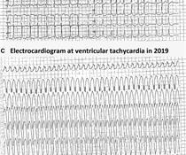
Electrocardiogram (ECG) showed sustained monomorphic VT at a rate of 160 bpm. This converted to normal sinus rhythm with intravenous amiodarone and metoprolol.
Players previously received an electrocardiogram (ECG) performed an average of 239 days before infection. Finally, Takotsubo cardiomyopathy should be considered in COVID-19 patients with acute cardiac failure since it exhibits a clinical picture resembling viral myocarditis.
Electrocardiograms (EKG or ECG): This diagnostic method effectively detects irregular heartbeats or arrhythmias by analyzing the electrical signals in the heart. It serves as an initial assessment, and an MRI may be considered if other signs of cardiomyopathy are present.
Electromechanical association: a subtle electrocardiogram artifact. I initially considered Takotsubo cardiomyopathy as the cause of the ECG in today’s case, for this middle-aged woman with new chest pain and dyspnea — because of the diffuse T wave inversion with obvious QTc prolongation. Journal of Electrocardiology. 2012;45(1):15-17.
Electromechanical association: a subtle electrocardiogram artifact. Arterial pulse tapping artifact [link] This online article references the article below by Emre Aslanger, a great guy who occasionally corresponds with me about ECGs. Aslanger E, Yalin K. Journal of Electrocardiology. 2012;45(1):15-17. doi:10.1016/j.jelectrocard.2010.12.162.
A Deep Neural Network learning algorithm outperforms a conventional algorithm for emergency department electrocardiogram interpretation. This ECG comes from Pierre Taboulet ( [link] /)( [link] ) an ECG whiz who codes a lot of ECGs for Cardiologs' Artificial Intelligence Deep Neural Network algorithm ( [link] ). What an honor.
Santos Most Cited Article – Reduction in Hospitalization and Increase in Mortality Due to Cardiovascular Diseases during the COVID-19 Pandemic in Brazil Authors: Paulo Garcia Normando, José de Arimatéia Araujo-Filho, Gabriela de Alcântara Fonseca, Rodrigo Elton Ferreira Rodrigues, Victor Agripino Oliveira, Ludhmila Abrahão Hajjar, André Luiz (..)
Signify Research has just released a deep-dive qualitative analysis of developments around the use of AI to analyze and interpret electrocardiograms (ECGs), one of the world’s most ubiquitous diagnostic tests for cardiac disease. Figure 1: The AI-ECG competitive vendor landscape New Horizons However, dramatic changes are afoot.
The diagnosis was made based on classic findings of inflammation on an electrocardiogram associated with acute chest pain. Events of cardiomyopathy or cardiac failure were reported by eight participants after the Novavax COVID-19 Vaccine, Adjuvanted (0.03%) and one participant after placebo (<0.01%).
Hence, despite the transformative impact of ART on reducing cardiomyopathy and heart failure in PLWH, concerns persist! Further, PIs are known for disturbances in lipid metabolism, and NNRTIs like efavirenz contribute to metabolic changes, highlighting the complexity of assessing CVD risk in PLWH.
A discrepancy was observed between electrocardiogram (ECG) results and echocardiography or cardiac magnetic resonance (CMR) in diagnosing LVH, emphasising the need for comprehensive cardiac imaging. Of 143 patients surveyed, only three with LVH underwent diagnostic testing for FD, all testing negative.
We organize all of the trending information in your field so you don't have to. Join thousands of users and stay up to date on the latest articles your peers are reading.
You know about us, now we want to get to know you!
Let's personalize your content
Let's get even more personalized
We recognize your account from another site in our network, please click 'Send Email' below to continue with verifying your account and setting a password.


Let's personalize your content