This site uses cookies to improve your experience. To help us insure we adhere to various privacy regulations, please select your country/region of residence. If you do not select a country, we will assume you are from the United States. Select your Cookie Settings or view our Privacy Policy and Terms of Use.
Cookie Settings
Cookies and similar technologies are used on this website for proper function of the website, for tracking performance analytics and for marketing purposes. We and some of our third-party providers may use cookie data for various purposes. Please review the cookie settings below and choose your preference.
Used for the proper function of the website
Used for monitoring website traffic and interactions
Cookie Settings
Cookies and similar technologies are used on this website for proper function of the website, for tracking performance analytics and for marketing purposes. We and some of our third-party providers may use cookie data for various purposes. Please review the cookie settings below and choose your preference.
Strictly Necessary: Used for the proper function of the website
Performance/Analytics: Used for monitoring website traffic and interactions
Her ED echo is diagnostic of apical ballooning, also known as "stress cardiomyopathy" (SCM) or "takostubo cardiomyopathy" (because the heart, with its apical ballooning, resembles the Japanese octopus trap called a "takostubo").
Echocardiograms using the robotic arm resulted in the same diagnosis as conventional in-person echocardiography in 98% of cases (papillary muscle level obstruction was missed in one case). tim.hodson Thu, 08/29/2024 - 11:39 Aug. 28, 2024 — New research presented at this year’s ESC Congress 2024 in London, UK (Aug. 30 – Sept.
Researchers used an AI-enabled digital stethoscope that captures electrocardiogram ( ECG ) data and heart sounds to identify twice as many cases of peripartum cardiomyopathy as compared to regular care, according to a news release from the American Heart Association.
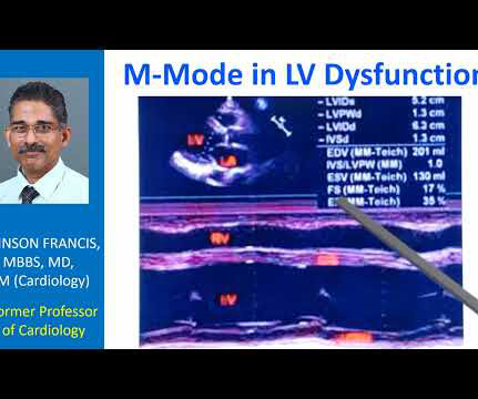
Transcript of the video: This is a still image of M-Mode Echocardiogram. Here it is 1 and in cases like asymmetric septal hypertrophy or hypertrophic cardiomyopathy, there will be disproportionate thickening of the septum. M-Mode is Time-Motion Mode. The horizontal axis is time. Vertical axis is distance from the transducer.
And of course Ken's comments at the bottom) An elderly obese woman with cardiomyopathy, Left bundle branch block, and chronic hypercapnea presented hypoxic with altered mental status. I do not see OMI here and all trops were only minimally elevated, consistent with either chronic injury from cardiomyopathy or with acute injury from sepsis.
BackgroundAbnormal substrate on invasive electroanatomic mapping (EAM) correlates with areas of myocardial thinning and fibrofatty replacement in Arrhythmogenic Cardiomyopathy (ACM). Transthoracic echocardiogram revealed normal biventricular function and dimension. Twelve-lead ECG showed diffuse low-voltage QRS complexes.
11, 2025 UltraSight, a company committed to enhancing the efficiency and productivity of cardiac ultrasound,recentlyannounced support from Bristol Myers Squibb (BMS) for a study that aims to improve access to echocardiographic assessments for patients with obstructive hypertrophic cardiomyopathy (oHCM). tim.hodson Tue, 02/18/2025 - 16:17 Feb.
Clinical introduction A patient in their 30s had been diagnosed with peripartum cardiomyopathy, pulmonary oedema, with severe left ventricular dysfunction at the seventh month of gestation in the third pregnancy in their late 20s. Echocardiogram, CT aortogram and late gadolinium imaging of the aorta have been shown in figure 1.
Example here: DIffuse ST Elevation with Apical Ballooning: is it Takotsubo Stress Cardiomyopathy? Stress induced cardiomyopathy (Takotsubo like LV dysfunction) possible. Outcome : She was diagnosed with stress cardiomyopathy, though it is not entirely classic. Therefore, the cath lab was activated.
BackgroundSigmoid Ventricular Septum (SVS) is a type of hypertrophic cardiomyopathy characterized by a reduced angle between the basal interventricular septum and the ascending aorta, and SVS can lead to dynamic Left Ventricular Outflow Tract obstruction (LVOTO) during hypercontractile states.
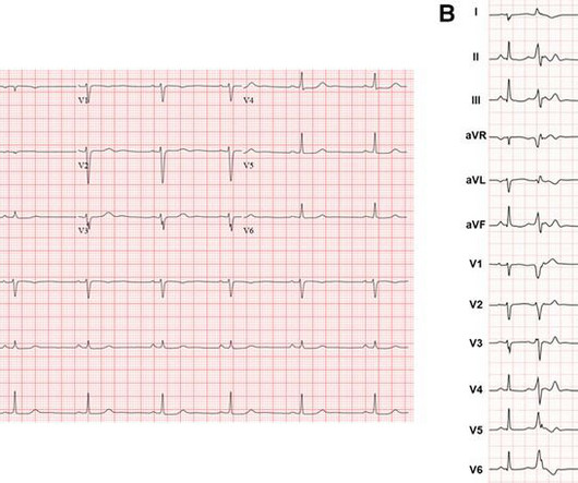
Echocardiogram showed an anteroapical wall motion abnormality. This is highly suspicious for acute anterior STEMI. However, she was found to have a fatal pontine hemorrhage and had a maximum troponin I, at 12 hours after presentation, of 2.0
This can manifest as a multitude of pathologies including left ventricular dysfunction, myocarditis, cardiomyopathy, accelerated atherosclerosis, and coronary vasospasm.
Hypertrophic cardiomyopathy is a genetic disorder with a guarded prognosis which occurs in about 1:500 individuals. The most common symptom of hypertrophic cardiomyopathy is dyspnoea which occurs in 90% of cases and is due to elevated left ventricular diastolic pressures as a consequence of the diastolic dysfunction.
Introduction:Mavacamten is a cardiac myosin inhibitor that has been approved for obstructive hypertrophic cardiomyopathy (oHCM). LVOT obstruction resolved on follow up echocardiogram. Circulation, Volume 150, Issue Suppl_1 , Page A4123391-A4123391, November 12, 2024.
Complete left bundle branch block (CLBBB)-like QRS morphology of right ventricular pacing at pacemaker implantation satisfying the American Heart Association/American College of Cardiology Foundation/Heart Rhythm Society criteria of CLBBB was associated with development of pacing induced cardiomyopathy.
Impact of preceding systolic heart failure on risk of pacemaker-induced cardiomyopathy Abstract Background Pacemaker-induced cardiomyopathy is a well described phenomenon in patients with preserved ejection fraction at the time of permanent pacemaker implant.
This comprehensive evaluation included the use of ultrasound echocardiograms, computed tomography (CT) scans, electrocardiograms, mutagenesis analysis, and structural analysis to gain insights into the patient's condition and the underlying mechanisms of PD. Further genetic testing identified a homozygous mutation c.2662G>T
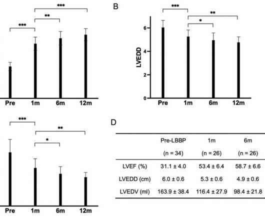
Patients with ischemic cardiomyopathy, left ventricular noncompaction, significant late gadolinium enhancement (LGE) on cardiac magnetic resonance imaging (CMR), and indications for an implantable cardioverter-defibrillator (ICD) as secondary prevention were excluded.ResultsPost-LBBP, the LVEF improved from 31.14.0% to 61.06.0% (P<0.001).
2. Baseline fibrotic substrate from dilated cardiomyopathy leading to VT. Corresponding echocardiogram demonstrated LV systolic dysfunction with an EF 30%. I interpreted the ECG as VT with two primary etiological possibilities: 1. Abrupt plaque ulceration of Type 1 ACS leading to VT.
In preparation for the ABIM Cardiovascular Disease exam, check out the BoardVitals Cardiology Board Review Question Bank and we’ll make sure you’re well versed in the following 13 areas covered on the exam: Multiple-Choice Component Arrhythmias 15% Coronary Artery Disease 23% Heart Failure and Cardiomyopathy 17% Valvular Disease 15% Pericardial (..)
Background:Mavacamten is the only cardiac myosin inhibitor approved by the US FDA for treatment of symptomatic New York Heart Association class II-III obstructive hypertrophic cardiomyopathy (HCM) patients. We report results from the mavacamten REMS database (28-Apr-2022 to 27-Feb-2024).Methods:Data were women; 64.6%
We explored the association with early CTRCD (new cardiomyopathy, heart failure, or left ventricular ejection fraction <50%), or left ventricular ejection fraction <40%, up to 12 months after treatment. positive screen), respectively. positive screen), respectively. interquartile range, 13.417.1] interquartile range, 13.417.1]
Examples of cardio embolic stroke etiology include: 1. Atrial Fibrillation 2. Cardiomyopathy with mural thrombus 3. Patent Foramen Ovale 4. Severe calcific Aortic (valve) Stenosis 5. Mechanical prosthetic valve Severe carotid artery stenosis is also implicated in embolic stroke. Here is the admission ECG.
So the artery had completely spontaneously reperfused prior to intervention; the duration of occlusion was perhaps 2 hours The troponin peaked at 60,000 ng/L (a very large infarction) Formal bubble contrast echocardiogram --The estimated left ventricular ejection fraction is 46%. He has "ischemic cardiomyopathy" and "congestive heart failure."
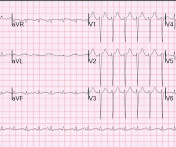
It turns out that she has hypertrophic cardiomyopathy. The diagnosis was a bit hard to find in the chart, and the echocardiogram did only stated "assymetric hypertrophy." Here is the echo report for this visit (after the negative angiogram): Hypertrophic cardiomyopathy with asymmetric septal hypertrophy.
Yes, COVID-19 symptoms can resemble a heart attack, including chest pain, shortness of breath, and changes in echocardiogram or EKG. Echocardiogram: Utilizing sound waves, this test generates images of the heart and its chambers to identify potential issues. Can COVID-19 symptoms mimic a heart attack?
Enlargement of the left ventricular cavity, increased wall thickness and increased trabeculations in athlete’s heart will have to be differentiated from conditions like dilated cardiomyopathy, hypertrophic cardiomyopathy and isolated left ventricular non-compaction. Differentiating Athlete’s Heart From Cardiomyopathies – The Left Side.
A 35-year-old gravida 1, para 0 with biventricular heart failure (LVEF 25%), nonischemic cardiomyopathy, history stroke, history of left ventricular thrombus, class III obesity, and chronic kidney disease who had been followed by Cardio-Obstetrics throughout her pregnancy presented at 34 weeks gestation for planned induction of labor.
A pre-procedural transesophageal echocardiogram showed a left ventricular (LV) ejection fraction of 55%, mild concentric LV hypertrophy, and mild left atrial enlargement. Despite alcohol cessation, he continued to have paroxysmal AF with severe fatigue and palpitations. These enlargements were new compared with imaging one year prior.
C ASE F ollow- U p: In view of this patient's CP at the time he was seen in the ED — the 2 slightly elevated troponin values — and the abnormal ST-T wave findings on the initial ECG ( with all of these ST-T wave changes being new since the baseline ECG done 9 years earlier ) — Cardiac cath was performed. Cardiac cath showed normal coronary arteries.
We describe a case of ventricular pacemaker spikes delivered on the T wave causing PMVT.Case:A 53-year-old female with CAD s/p stent, postpartum cardiomyopathy s/p Bi-V CRT-D (Boston Scientific G124), and paroxysmal atrial fibrillation presented for elective endoscopy and colonoscopy to evaluate her dysphagia and abdominal pain.
The combination of prolonged QT and deep T wave inversion throughout the precordium is typical of Takotsubo syndrome, or Stress Cardiomyopathy – which can occur in the context of a physiologically distressed ICU patient, further compromising their hemodynamics. The coronary angiogram revealed no critical stenosis, or acute plaque ulceration.
by making it clear to everyone that this is NOT an EKG that one sees with takotsubo cardiomyopathy. Hospital Course The patient was taken emergently to the cath lab which did not reveal any significant coronary artery disease, but she was noted to have reduced EF consistent with Takotsubo cardiomyopathy.
His echocardiogram showed normal wall motion. The 3 most common causes of ACS ( A cute C oronary S yndrome ) wit hout evidence of obstructive coronary disease on cath are: i ) Myocarditis ( up to 1/3 of these patients ); ii ) Takotsubo cardiomyopathy; and , iii ) MINOCA. There are no further EKGs or troponin measurements.
So cardiomyopathies, valve problems, myocarditis and previous heart attacks all cause a problem with the pumping function of the heart. Overall though a normal cardiac MRI is even more reassuring than a normal echocardiogram. This is an ultrasound (a bit like the type that we use on pregnant women to look at the baby).
I have ordered an echocardiogram which will be done today, after that patient can be discharged to home with follow-up in 2 to 3 months." The echo was normal. Learning points 1. In this regular wide complex tachycardia , since the rhythm converted w adenosine, it is almost certainly SVT w aberrancy, which can be either: A.
These are reperfusion T-waves (the same thing as Wellens' waves) Echocardiogram Regional wall motion abnormality-distal septum and apex. Troponins may be negative with very rapid reperfusion, or measured too late, or chronically elevated due to cardiomyopathy or renal failure. Regional wall motion abnormality-distal inferior wall.
CardiomyopathyCardiomyopathy is a condition that affects the heart muscle, causing it to become enlarged, thick, or rigid. Excessive Alcohol or Drug Use Long-term abuse of alcohol or certain drugs can weaken the heart muscle, resulting in cardiomyopathy and eventually cardiomegaly.
Cardiomyopathies: These diseases affect the heart muscle, impairing its ability to pump blood effectively. Mutations in specific genes often cause hypertrophic cardiomyopathy and dilated cardiomyopathy. Heart imaging, such as echocardiograms or CT scans. Key screenings include: Blood pressure and cholesterol checks.
Objectives To describe a cohort of patients with arrhythmogenic left ventricular cardiomyopathy (ALVC), focusing on the spectrum of the clinical presentations. Twenty-one (41%) had normal echocardiogram, 13 (25%) a hypokinetic non-dilated cardiomyopathy (HNDC) and 17 (33%) a dilated cardiomyopathy (DCM).
4 Demonstrated Impact in Pregnant Women: A clinical study led by Mayo Clinic involving nearly 1,200 pregnant women in Nigeria highlighted the AI's effectiveness, identifying twice as many cases of pregnancy-related cardiomyopathy than standard care, with an impressive AUROC of 0.98, 100.0% for detection of LVEF below 40%, 84.8% Adedinsewo DA.
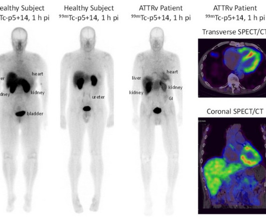
Blood was collected to assess serum biomarkers, and a transthoracic echocardiogram was performed. Patients with amyloid cardiomyopathy had significant 99mTc-p5+14 uptake in the heart, whereas no cardiac uptake was observed in healthy subjects. to assess safety and efficacy in patients with cardiac amyloidosis and healthy subjects.
Whether you’re gearing up for your Family Medicine or Cardiology Board Exam, you’ll need to master the topic of Hypertrophic Cardiomyopathy (HCM). Echocardiogram is indicated (Correct) C. Start aspirin and Plavix Correct answer: (B) (B) Echocardiogram is indicated. Hypertrophic cardiomyopathy is one of them.
We organize all of the trending information in your field so you don't have to. Join thousands of users and stay up to date on the latest articles your peers are reading.
You know about us, now we want to get to know you!
Let's personalize your content
Let's get even more personalized
We recognize your account from another site in our network, please click 'Send Email' below to continue with verifying your account and setting a password.


Let's personalize your content