This site uses cookies to improve your experience. To help us insure we adhere to various privacy regulations, please select your country/region of residence. If you do not select a country, we will assume you are from the United States. Select your Cookie Settings or view our Privacy Policy and Terms of Use.
Cookie Settings
Cookies and similar technologies are used on this website for proper function of the website, for tracking performance analytics and for marketing purposes. We and some of our third-party providers may use cookie data for various purposes. Please review the cookie settings below and choose your preference.
Used for the proper function of the website
Used for monitoring website traffic and interactions
Cookie Settings
Cookies and similar technologies are used on this website for proper function of the website, for tracking performance analytics and for marketing purposes. We and some of our third-party providers may use cookie data for various purposes. Please review the cookie settings below and choose your preference.
Strictly Necessary: Used for the proper function of the website
Performance/Analytics: Used for monitoring website traffic and interactions
What is the preferred order of vasopressors and ionotropes in the management of cardiogenicshock? How can we best pick up occult cardiogenicshock before it floured shock kicks in? The post Ep 164 CardiogenicShock Simplified appeared first on Emergency Medicine Cases.
Taking a step back , remember that sinus tachycardia is less commonly seen in OMI (except in cases of impending cardiogenicshock). Answer : Bedside ultrasound! Smith : RV infarct may also have this appearance on ultrasound. So hypoxia without B lines on lung ultrasound strongly weights toward PE. Both were wrong.
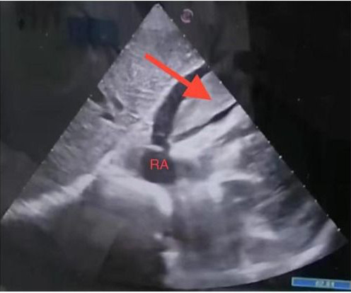
This paper reports a case of an elderly female patient who experienced severe chest pain and syncope during acupuncture therapy, subsequently diagnosed with traumatic hemopericardium and acute cardiac tamponade, complicated by cardiogenicshock. Under ultrasound guidance, pericardial puncture and drainage were successfully performed.
ACS and STEMI generally do not cause tachycardia unless there is cardiogenicshock. Then ACS (STEMI) might be primary; this might be cardiogenicshock. Even if this ECG is the first thing one sees (as it was for me), one should stop and think: "This is an unusual STEMI." Are the lungs clear? Is the patient cool and pale?
Background Pompe disease (PD) is a rare, progressive autosomal recessive lysosomal storage disorder that directly impacts mitochondrial function, leading to structural abnormalities and potentially culminating in heart failure or cardiogenicshock.
Smith comment: This patient did not have a bedside ultrasound. Had one been done, it would have shown a feature that is apparent on this ultrasound (however, this patient's LV function would not be as good as in this clip): This is recorded with the LV on the right. In fact, bedside ultrasound might even find severe aortic stenosis.
ET Main Tent (Hall B1) Self-expanding Versus Balloon-expandable Transcatheter Aortic Valve Replacement in Patients with Small Aortic Annuli: Primary Outcomes from the Randomized Smart Trial Effect of Edetate Disodium Based Chelation Infusions on Cardiovascular Events in Post-MI Patients with Diabetes: The TACT2 Trial Long-term Beta-blocker Treatment (..)
Case continued A bedside ultrasound showed diminished LV EF and of course bradycardia. RVMI explains part of the shock. If it did not do so, norepinephrine would be indicated to maintain systolic BP As for the escape rhythm: It is narrow complex, and therefore it is in the His bundle or above (not below the His bundle).
Thirty minutes later the first Troponin I came back elevated at 650 ng/L (normal <26), and bedside ultrasound found anteroseptal akinesia. But by this time the patient went into cardiogenicshock and passed away. But it didn’t meet traditional Sgarbossa criteria so the cath lab was not activated.
I would do bedside ultrasound to look at the RV, look for B lines as a cause of hypoxia (which would support OMI, and argue against PE), and if any doubt persists, a rapid CT pulmonary angiogram. As for the ECG, it could represent OMI, but RBBB is also a clue that it may be PE. There is sinus tachycardia at ~100/minute.
I have always said that tachycardia should argue against acute MI unless there is cardiogenicshock or 2 simultaneous pathologies. Sinus tachycardia, which exaggerates ST segments and implies that there is another pathology. We showed this in a recent analysis of UTROPIA data (see abstract below).
Just prior to transport, the patient became confused and agitated and, although blood pressure and pulse were OK, I was worried about cardiogenicshock. Diagnosis : Posterior MI, right? We intubated him. Cath lab The BP was 70/40 on arrival to the cath lab and received a balloon pump and norepinephrine.
Often, intravascular ultrasound or intravascular optical coherence tomography is requeried to make the diagnosis. were pretty sick, with mostly LM/pLAD lesions and high rates of cardiogenicshock. Type 2 is more difficult to appreciate on angiography than type 1. Lobo et al. where more than 3/4 of cases were NSTEMI).
An elderly man with sudden cardiogenicshock, diffuse ST depressions, and STE in aVR Literature 1. Widespread ST-depression with reciprocal aVR ST-elevation can be cause by: Heart rate related: tachyarrhythmia (e.g., A emergent cardiology consult can be helpful for equivocal cases. Left main? 3-vessel disease? Knotts et al.
Shocked x 2 without effect. Pads were placed with ultrasound guidance, so they were in the correct position. No adenosine was given (if you believe it is SVT, this is worth a try). However, this is not SVT. If it is VT, adenosine is safe but not effective. What to do now? If you believe it is SVT, then try adenosine. Patient intubated.
The patient in today’s case presented in cardiogenicshock from proximal LAD occlusion, in conjunction with a subtotally stenosed LMCA. Another approach is sympathetic chain (stellate ganglion) blockade if you have the skills to do it: it requires some expertise and ultrasound guidance. RCA — 100% proximal occlussion.
Case Continued 2 days later the patient became increasingly tachycardic, hypotensive, ashen, clammy (in cardiogenicshock) and had a new murmur. Rupture can be either free wall rupture (causing tamonade) or septal rupture, causing ventricular septal defect with left to right flow and resulting pulmonary edema and shock.
A bedside cardiac ultrasound was normal, with no effusion. Assessment was severe sudden cardiogenicshock. Clinically — the patient was felt to be in cardiogenicshock. This sinus tachycardia ( at ~130/minute ) — is consistent with the patient’s worsening clinical condition, with development of cardiogenicshock.
Why is the patient in shock? He was in profound cardiogenicshock. They did not have an ultrasound on the ambulance (some local crews are starting to utilize POC limited US in our service areas). There is an obvious inferior STEMI, but what else? This STE is diagnostic of Right Ventricular STEMI (RV MI).
Tachycardia is unusual for OMI, unless the patient is in cardiogenicshock (or getting close). A bedside ultrasound should be done to assess volume and other etiologies of tachycardia, but if no cause of type 2 MI is found, the cath lab should be activated NOW. We can see enough to make out that the rhythm is sinus tachycardia.
On arrival in the ED, a bedside ultrasound showed poor LV function (as predicted by the Queen of Hearts) with diffuse B-lines. I don't know what the device algorithm interpretation stated. I am not certain if there was a prehospital cath lab activation, but there should be. Initial BP was 120/96, HR 102, SpO2 98%.
Whenever there is tachycardia, I am skeptical of OMI unless it has led to severely compromised ejection fracction with cardiogenicshock. I suspect pulmonary edema, but we are not given information on presence of B-lines on bedside ultrasound, or CXR findings. Or I suspect that there is OMI simultaneous with another pathology.
The post EM Quick Hits 28 CardiogenicShock, Radiation Dose in Pregnancy, PoCUS in Airway Management, VIPIT, Angiotensin II, Short-Term Steroid Safety appeared first on Emergency Medicine Cases.
We organize all of the trending information in your field so you don't have to. Join thousands of users and stay up to date on the latest articles your peers are reading.
You know about us, now we want to get to know you!
Let's personalize your content
Let's get even more personalized
We recognize your account from another site in our network, please click 'Send Email' below to continue with verifying your account and setting a password.


Let's personalize your content