This site uses cookies to improve your experience. To help us insure we adhere to various privacy regulations, please select your country/region of residence. If you do not select a country, we will assume you are from the United States. Select your Cookie Settings or view our Privacy Policy and Terms of Use.
Cookie Settings
Cookies and similar technologies are used on this website for proper function of the website, for tracking performance analytics and for marketing purposes. We and some of our third-party providers may use cookie data for various purposes. Please review the cookie settings below and choose your preference.
Used for the proper function of the website
Used for monitoring website traffic and interactions
Cookie Settings
Cookies and similar technologies are used on this website for proper function of the website, for tracking performance analytics and for marketing purposes. We and some of our third-party providers may use cookie data for various purposes. Please review the cookie settings below and choose your preference.
Strictly Necessary: Used for the proper function of the website
Performance/Analytics: Used for monitoring website traffic and interactions
This is a value typical for a large subacute MI, n ormal value 48 hours after myocardial infarction is associated with Post-Infarction Regional Pericarditis ( PIRP ). As already mentioned, this patient could have post-infarction regional pericarditis from a large completed MI. The VSR is what is causing the cardiogenicshock!
A previously healthy 53 yo woman was transferred to a receiving hospital in cardiogenicshock. Well, don't we see diffuse ST Elevation in Myo-pericarditis (with STD in aVR)? Referring to Figure-1 — this 53-year old woman who presented in extremis with cardiogenicshock and an initial pH = 6.9, and K was normal.
I have always said that tachycardia should argue against acute MI unless there is cardiogenicshock or 2 simultaneous pathologies. PR depression, which suggests pericarditis 4. We also showed that, of 47 cases of pericarditis with ST elevation, none had ST depression in aVL. ) Absence of any ST depression in aVL. (
When there is MI extending all the way to the epicardium (transmural), that infarcted epicardium is often inflamed (postinfarction regional pericarditis, or PIRP). What complication is the patient with post-infarction regional pericarditis at risk for? The initial troponin I was 23.7 This was the 12-lead ECG. 3) Oliva et al. (4)
The patient died of cardiogenicshock within 24 hours despite mechanical circulatory support. The axis is to the right and QRS complexes in lead I and aVL are predominantly negative suggesting LPFB. This patient at cath had a large CX occlusion with a massive troponin release. Troponin T >42.000ng/L.
Tachycardia is unusual for OMI, unless the patient is in cardiogenicshock (or getting close). The "flu-like" illness suggests myo- or pericarditis, but that would be a diagnosis of exclusion. The ECG has a lot of artifact, and the amplitude is very small, making interpretation challenging. The case continues.
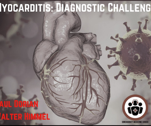
When should we consider myocarditis or pericarditis in patients with recent COVID-19 infection or COVID mRNA vaccination, and which of these patients require workups? Does a negative high sensitivity troponin or CRP rule out myocarditis? What is the role of PoCUS in the diagnosis and prognosis of myocarditis?
We organize all of the trending information in your field so you don't have to. Join thousands of users and stay up to date on the latest articles your peers are reading.
You know about us, now we want to get to know you!
Let's personalize your content
Let's get even more personalized
We recognize your account from another site in our network, please click 'Send Email' below to continue with verifying your account and setting a password.


Let's personalize your content