This site uses cookies to improve your experience. To help us insure we adhere to various privacy regulations, please select your country/region of residence. If you do not select a country, we will assume you are from the United States. Select your Cookie Settings or view our Privacy Policy and Terms of Use.
Cookie Settings
Cookies and similar technologies are used on this website for proper function of the website, for tracking performance analytics and for marketing purposes. We and some of our third-party providers may use cookie data for various purposes. Please review the cookie settings below and choose your preference.
Used for the proper function of the website
Used for monitoring website traffic and interactions
Cookie Settings
Cookies and similar technologies are used on this website for proper function of the website, for tracking performance analytics and for marketing purposes. We and some of our third-party providers may use cookie data for various purposes. Please review the cookie settings below and choose your preference.
Strictly Necessary: Used for the proper function of the website
Performance/Analytics: Used for monitoring website traffic and interactions
Cardiogenicshock (CS)is the most feared event following STEMI. We tend to perceive CS as an exclusive complication of STEMI. The incidence is half of that of STEMI, i.e., 2.5-5%. might show little elevation with considerable overlap of left main STEMI vs NSTEMI ) 2.Onset How is CS in NSTEMI different ?
The paramedic called the EM physician ahead of arrival and discussed the case and ECGs, and both agreed upon activating "Code STEMI" (even though of course it is not STEMI by definition), so that the acute LAD occlusion could be treated as fast as possible. Long term outcome is unavailable. So the cath lab was activated.
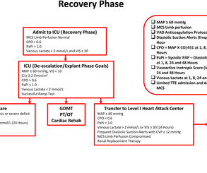
Introduction Cardiogenicshock (CS) complicates 5%–15% of cases of acute myocardial infarction (AMI) with inpatient mortality greater than 40%. The implementation of standardised protocols may improve clinical outcomes in patients with AMI-CS.
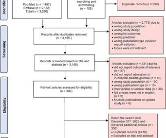
Background Hyperglycemia, characterized by elevated blood glucose levels, is frequently observed in patients with acute coronary syndrome, including ST-elevation myocardial infarction (STEMI). There are conflicting sources regarding the relationship between hyperglycemia and outcomes in STEMI patients. 3.45) and 4.47 (95% CI: 2.54–7.87),
Now appears to be in cardiogenicshock." However, cardiogenicshock usually takes some time to develop, so it is probably subacute." This can only be due to STEMI. Cardiogenicshock and ACS is an indication for the cath lab, even if you don't think there is OMI. I was texted these ECGs.
Thus, this is BOTH an anterior and inferior STEMI in the setting of RBBB. How old is this antero-inferior STEMI? Although acute anterior STEMI frequently has narrow QR-waves within one hour of onset (1. Although acute anterior STEMI frequently has narrow QR-waves within one hour of onset (1. Could it be acute (vs.
As a low-volume PCI centre in the Middle East, we wanted to find out if the outcomes of our PCI procedures are different from those of high-volume PCI centres in the UK and the Western world. Prospectively collected data of all comers for PCI (urgent and elective) were retrospectively analysed. and the average number of stents 2.6.
Then the notes mention "cardiogenicshock" but without any reference to a cardiac echo or to a chest x-ray. Cardiologist note says: "Elevated troponin explained by type II MI due to her shock." Trop T now very high, well into the range one sees with a STEMI; very unusual in type II MI. Was there pulmonary edema?
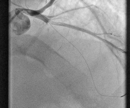
Troponin T peaked at 2074 ng/L (very high, typical of OMI/STEMI). Post PCI the patient became gravely hypotensive and "shocky". She stabilized on dobutamine and levosimendan infusions that could be discontinued after 24 hours. The tricuspid annular plane systolic excursion (TAPSE) improved from 15mm to 19mm within the first 24 hours.
There is an obvious inferior posterior STEMI(+) OMI. Methods Retrospective study of consecutive inferior STEMI , comparing ECGs of patients with, to those without, RVMI, as determined by angiographic coronary occlusion proximal to the RV marginal branch. What is the atrial activity? Is it sinus arrest with junctional escape?
The ECG was read as "No STEMI" and the patient was treated like an average chest pain patient (despite the fact that a chest pain patient with active pain and active subendocardial ischemia is very high risk). The notes now refer to the patient being in cardiogenicshock, on pressors.
Code STEMI was activated by the ED physician based on the diagnostic ECG for LAD OMI in ventricular paced rhythm. This was several months after the 2022 ACC Guidelines adding modified Sgarbossa criteria as a STEMI equivalent in ventricular paced rhythm). LAFB, atrial flutter, anterolateral STEMI(+) OMI. Limkakeng AT.
When total LM occlusion does present with STE in aVR, there is ALWAYS ST Elevation elsewhere which makes STEMI obvious; in other words, STE is never limited to only aVR but instead it is part of a massive and usually obvious STEMI. All are, however, clearly massive STEMI. This is her ECG: An obvious STEMI, but which artery?
This has been termed a “STEMI equivalent” and included in STEMI guidelines, suggesting this patient should receive dual anti-platelets, heparin and immediate cath lab activation–or thrombolysis in centres where cath lab is not available. aVR ST segment elevation: acute STEMI or not? aVR ST Segment Elevation: Acute STEMI or Not?
Assessment was severe sudden cardiogenicshock. However, in multiple studies, even in the absence of AMI, both acute and chronic myocardial injury (as diagnosed by any elevated cTn) are powerful markers of adverse outcomes in both the short and long term. 12 All STEMI patients had very high cTn typical of STEMI (cTnT > 1.0
He was in cardiogenicshock requiring an impella for several days after cath. Sooner identification likely leads to better outcomes, and in this case may have allowed prevention of cardiac arrest and better long-term outcome. No further troponins were measured. Such is the situation in today's post by Drs.
A recent study found that SCAD causes almost 20% of STEMI in young women. examined SCAD presenting as STEMI (unlike Hassan et al. were pretty sick, with mostly LM/pLAD lesions and high rates of cardiogenicshock. I had no idea SCAD was so common a cause of acute STEMI in younger women, even when they are non-smokers.
The axiom of "type 1 (ACS, plaque rupture) STEMIs are not tachycardic unless they are in cardiogenicshock" is not applicable outside of sinus rhythm. Is that an obvious STEMI underneath that rhythm? Again, not an expected outcome with diltiazem). Is this inferor STEMI? Atrial Flutter with Inferior STEMI?
PCI mid LCx So this is an OMI (Occlusion Myocardial Infarction), but not a STEMI Echo: Decreased left ventricular systolic performance, mild/moderate. The patient went into cardiogenicshock and ultimately died of this MI. Angiogram: LM 30% ostial. LAD 80% mid LCx occluded mid (acute infarct lesion) RCA 80% mid. Sandoval Y.
Whenever there is tachycardia, I am skeptical of OMI unless it has led to severely compromised ejection fracction with cardiogenicshock. Supply-demand mismatch can cause ST Elevation (Type 2 STEMI). Also see these posts of Type II STEMI. Truly, the Marquette 12 SL algorithm correctly identifies this STEMI.
We organize all of the trending information in your field so you don't have to. Join thousands of users and stay up to date on the latest articles your peers are reading.
You know about us, now we want to get to know you!
Let's personalize your content
Let's get even more personalized
We recognize your account from another site in our network, please click 'Send Email' below to continue with verifying your account and setting a password.


Let's personalize your content