This site uses cookies to improve your experience. To help us insure we adhere to various privacy regulations, please select your country/region of residence. If you do not select a country, we will assume you are from the United States. Select your Cookie Settings or view our Privacy Policy and Terms of Use.
Cookie Settings
Cookies and similar technologies are used on this website for proper function of the website, for tracking performance analytics and for marketing purposes. We and some of our third-party providers may use cookie data for various purposes. Please review the cookie settings below and choose your preference.
Used for the proper function of the website
Used for monitoring website traffic and interactions
Cookie Settings
Cookies and similar technologies are used on this website for proper function of the website, for tracking performance analytics and for marketing purposes. We and some of our third-party providers may use cookie data for various purposes. Please review the cookie settings below and choose your preference.
Strictly Necessary: Used for the proper function of the website
Performance/Analytics: Used for monitoring website traffic and interactions
A 20-something presented after a huge verapamil overdose in cardiogenicshock. And she does not know that this is an overdose; she thinks it is a patient with chestpain!! Today's patient is a young male who presented in cardiogenicshock following a massive verapamil overdose. The initial K was 3.0
Written by Jesse McLaren Two patients in their 70s presented to the ED with chestpain and RBBB. Patient 1 : a 75 year old called paramedics with one day of left shoulder pain which migrated to the central chest, which was worse with deep breaths. Do either, both, or neither have occlusion MI? Vitals were normal.
Bad chest pressure with severe left shoulder pain 3 nights ago. Now appears to be in cardiogenicshock." However, cardiogenicshock usually takes some time to develop, so it is probably subacute." Cardiogenicshock and ACS is an indication for the cath lab, even if you don't think there is OMI.
A 50-something man presented in shock with severe chestpain. The patient was in clinical shock with a lactate of 8. His prehospital ECG was diagnostic of inferior posterior OMI. BP was 108 systolic (if a cuff pressure can be trusted) but appeared to be maintaining BP only by very high systemic vascular resistance.
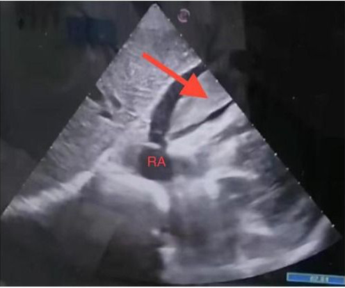
The VSR is what is causing the cardiogenicshock! Mechanical complications occur acutely and significantly alter hemodynamics leading to comp ensatory mechanism which usually involve vasoconstriction and tachycardia, both hallmarks of cardiogenicshock. PIRP is strongly associated with myocardial rupture.
A middle-aged patient with lung cancer had presented to clinic complaining of generalized malaise, cough, and chestpain. Symptoms other than chestpain (malaise, cough in a cancer patient) 2. I have always said that tachycardia should argue against acute MI unless there is cardiogenicshock or 2 simultaneous pathologies.
[link] A 30 year-old woman was brought to the ED with chestpain. She had given birth a week ago, and she had similar chestpain during her labor. She attributed the chestpain to anxiety and stress, saying "I'm just an anxious person." Lobo et al. examined SCAD presenting as STEMI (unlike Hassan et al.
Case submitted by Rachel Plate MD, written by Pendell Meyers A man in his 70s presented with chestpain which had started acutely at rest and has lasted for 2 hours. The pain was still ongoing at arrival. He was in cardiogenicshock requiring an impella for several days after cath.
This paper reports a case of an elderly female patient who experienced severe chestpain and syncope during acupuncture therapy, subsequently diagnosed with traumatic hemopericardium and acute cardiac tamponade, complicated by cardiogenicshock.
This 54 year old patient with a history of kidney transplant with poor transplant function had been vomiting all day when at 10 PM he developed severe substernal crushing chestpain. ACS and STEMI generally do not cause tachycardia unless there is cardiogenicshock. He had this ECG recorded. Are the lungs clear?
Taking a step back , remember that sinus tachycardia is less commonly seen in OMI (except in cases of impending cardiogenicshock). But the History in today's case was acute shortness of breath with dizziness and lightheadedness — and, essentially without chestpain! Additionally, there is borderline right axis deviation.
A man in his 70s with past medical history of hypertension, dyslipidemia, CAD s/p left circumflex stent 2 years prior presented to the ED with worsening intermittent exertional chestpain relieved by rest. This episode of chestpain began 3 hours ago and was persistent even at rest. Troponin was ordered.
A 56 yo f with h/o HTN and hypercholesterolemia called EMS from home after onset of L chestpain radiating to the left arm. She arrived comatose and in cardiogenicshock and the following ECG was recorded. Before EMS arrived, she had "seizure activity" and became unresponsive. She was intubated.
Jesse McLaren (@ECGcases), of Emergency Medicine Cases Reviewed by Pendell Meyers and Steve Smith An 85yo with a history of hypertension developed chestpain and collapsed, and had bystander CPR. On arrival, GCS was 13 and the patient complained of ongoing chestpain. Vitals were HR 58 BP 167/70 R20 sat 96%.
Just the fact of chestpain and highly elevated troponin is enough to activate the cath lab, but here you can see just how subtle hyperacute T-waves can be. 2) Typical persistent chestpain with a sigificantly elevated troponin is OMI until proven otherwise, regardless of the ECG.
PEARL # 2: In the absence of associated heart failure ( cardiogenicshock ) — sinus tachycardia is not a common finding in acute MI. Today’s patient presented to the ED not only with chestpain — but also with shortness of breath , therefore with a history potentially consistent with the diagnosis.
Subsequently, he developed chestpain with hypotension, diffuse ST elevations on ECG, and hsTropI of 638 ng/L. L/min/m2, suggestive of myopericarditis with cardiogenicshock. Microbiological workup and abdominal imaging were unremarkable. IABP was inserted.
It was edited by Smith CASE : A 52-year-old male with a past medical history of hypertension and COPD summoned EMS with complaints of chestpain, weakness and nausea. Authors' commentary: Cardiogenicshock in the setting of severe aortic stenosis. Fundamentally, cardiogenicshock is an issue of decreased cardiac output.
ET Main Tent (Hall B1) Self-expanding Versus Balloon-expandable Transcatheter Aortic Valve Replacement in Patients with Small Aortic Annuli: Primary Outcomes from the Randomized Smart Trial Effect of Edetate Disodium Based Chelation Infusions on Cardiovascular Events in Post-MI Patients with Diabetes: The TACT2 Trial Long-term Beta-blocker Treatment (..)
This is one case where it made a difference: Right Ventricular MI seen on ECG helps Angiographer to find Culprit Lesion Nevertheless, it is sometimes a fun academic exercise to try to predict the infarct artery: An elderly patient had onset of chestpain one hour prior. His included cardiogenicshock, V Tach, AV block.
A middle aged man had off and on chestpain for 2 weeks, then 2 hours of more severe and constant pain. Just prior to transport, the patient became confused and agitated and, although blood pressure and pulse were OK, I was worried about cardiogenicshock. He did not get prehospital activation. What do you think?
He went into cardiogenicshock and is intubated in the cardiac ICU. Cortland : Thank you so much for your reply! I just got the follow up that he had a near complete very proximal LAD occlusion , and a complete PDA occlusion. Not the culprit artery I was expecting but potentially a wraparound LAD?
Wellens' is a syndrome of a painless period following an anginal (chestpain) event. When there is tachycardia, the patient is in cardiogenicshock with very poor LV function on bedside echo. We hope you can see the difference: See these cases for more examples: Syncope, Shock, AV block, Large RV, "Anterior" ST Elevation.
Then the notes mention "cardiogenicshock" but without any reference to a cardiac echo or to a chest x-ray. Now chestpain free. Cardiologist note says: "Elevated troponin explained by type II MI due to her shock." They were worried that the syncope was seizure and that she had brain mets.
And some similar ECGs from Pulmonary Embolism: A young woman with altered mental status and hypotension An elderly woman transferred to you for chestpain, shortness of breath, and positive troponin - does she need the cath lab now? Tachycardia is unusual in ACS unless there is cardiogenicshock or a second simultaneous pathology.
This was my response: If it is the right clinical situation, such as acute chest discomfort, it looks like proximal left anterior descending occlusion with right bundle branch block and left anterior fascicular block. Because of the tachcardia, I would expect her to be very poor left ventricular function and maybe Cardiogenicshock.
The best course is to wait until the anatomy is defined by angio, then if proceeding to PCI, add Cangrelor (an IV P2Y12 inhibitor) I sent the ECG and clinical information of a 90-year old with chestpain to Dr. McLaren. An elderly man with sudden cardiogenicshock, diffuse ST depressions, and STE in aVR Literature 1.
But the symptoms returned with similar pattern – provoked by exertion, and alleviated with rest; except that on each occasion the chestpain was a little more intense, and the needed recovery period was longer in duration. Then, she attempted to reengage the activities at hand, and initially tolerated this well.
Written by Pendell Meyers A man in his 40s called EMS for acute chestpain that awoke him from sleep, along with nausea and shortness of breath. Smith : LAD OMI with RBBB/LAFB is not only subtle on the ECG, but most of these patients are extremely ill: most I have seen are post-ROSC, in cardiogenicshock, or arrested shortly after.
He woke up alert and with chestpain which he also had experienced intermittently over the previous few days. The history in today's case with sudden loss of consciousness followed by chestpain is very suggestive of ACS and type I ischemia as the cause of the ECG changes. What do you think? This is an ominous sign.
They had difficulty describing their symptoms, but complained of severe weakness, nausea, vomiting, headache, and chestpain. They described the chestpain as severe, crushing, and non-radiating. Tachycardia is unusual for OMI, unless the patient is in cardiogenicshock (or getting close).
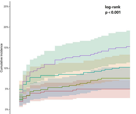
ObjectiveAlthough the association between admission glucose (AG) and major adverse cardiac events (MACE) is well-documented, its relationship with 30-day MACE in patients presenting with cardiac chestpain remains unclarified.
The following are the KEY clinical and ECG features that establish the diagnosis of W ellens ' S yndrome : There should be a history of prior chestpain that has resolved at the time the defining ECG is obtained. The ChestPain required for the definition of Wellens' Syndrome occurred at the time of coronary occlusion.
Edits by Meyers and Smith A man in his 70s with PMH of hypertension, hyperlipidemia, type 2 diabetes, CVA, dual-chamber Medtronic pacemaker, presented to the ED for evaluation of acute chestpain. Triage ECG: What do you think? This is diagnostic of proximal LAD occlusion. This is a huge anterolateral OMI. I cannot be anything else.
He had concurrent sharp substernal chestpain that resolved, but palpitations continued. Over past 3 months, he has had similar intermittent episodes of sharp chestpain while running, but none at rest. A 50-something presented with s udden onset palpitations 8 hrs prior while sitting at desk at work. Patient intubated.
Some patients have baseline RBBB with LAFB, but in patients with likely ACS, these are associated with severe infarction with cardiac arrest, cardiogenicshock or impending shock. Here are some cases of RBBB with LAFB: What is the Diagnosis in this 70-something with ChestPain?
The patient’s chestpain spontaneously resolved before he was evaluated and has a repeat ECG obtained at 22:12 obtained shown below. In context, of course, it is clear that the patient is reperfusing, as pain has dissipated and the diagnostic findings of OMI have become more nonspecific. This ECG is more difficult.
This 51 yo male complained of chestpain, then had a v fib arrest. He was in cardiogenicshock. The patient died 8 hours later of cardiogenicshock. Here are more examples of wide complex tachycardia: these are all a mix of ventricular tachycardia and SVT with aberrancy.
All of the patients presented with chestpain , and they are all in triage. The patient died of cardiogenicshock within 24 hours despite mechanical circulatory support. Triage is backed up, and 10 minutes into your shift one of the ED nurses brings your several ECG s that has not been overread by a physician.
The patient in today’s case is a previously healthy 40-something male who contacted EMS due to acute onset crushing chestpain. The pain was 10/10 in intensity radiating bilaterally to the shoulders and also to the left arm and neck. Written By Magnus Nossen — with edits by Ken Grauer and Smith. The below ECG was recorded.
This pattern is essentially always accompanied by cardiogenicshock and high rates of VT/VF arrest, etc. The patient arrived to the ED in cardiogenicshock but awake. What is the Diagnosis in this 70-something with ChestPain? 68 minutes with chest compressions, full recovery.
He was asked multiple times about chestpain or dyspnea, but repeatedly denied any such symptoms. Patient denied chestpain on initial review of symptoms. Was now endorsing chestpain which began 30 minutes ago. Upon further questioning, he states that he has had intermittent chestpain since yesterday.
A man in his 60's presented after 4 days of chestpain, with some increase of pain on the day of presentation. Exact pain history was difficult to ascertain. Case Continued 2 days later the patient became increasingly tachycardic, hypotensive, ashen, clammy (in cardiogenicshock) and had a new murmur.
His comments/questions are inserted below the ECG: A 50-something woman presented with 3 days of intermittent chestpain that became worse on the day of presentation, with diaphoresis and radiation to the left arm, as well as abdominal pain. This is her ECG: An obvious STEMI, but which artery?
We organize all of the trending information in your field so you don't have to. Join thousands of users and stay up to date on the latest articles your peers are reading.
You know about us, now we want to get to know you!
Let's personalize your content
Let's get even more personalized
We recognize your account from another site in our network, please click 'Send Email' below to continue with verifying your account and setting a password.


Let's personalize your content