This site uses cookies to improve your experience. To help us insure we adhere to various privacy regulations, please select your country/region of residence. If you do not select a country, we will assume you are from the United States. Select your Cookie Settings or view our Privacy Policy and Terms of Use.
Cookie Settings
Cookies and similar technologies are used on this website for proper function of the website, for tracking performance analytics and for marketing purposes. We and some of our third-party providers may use cookie data for various purposes. Please review the cookie settings below and choose your preference.
Used for the proper function of the website
Used for monitoring website traffic and interactions
Cookie Settings
Cookies and similar technologies are used on this website for proper function of the website, for tracking performance analytics and for marketing purposes. We and some of our third-party providers may use cookie data for various purposes. Please review the cookie settings below and choose your preference.
Strictly Necessary: Used for the proper function of the website
Performance/Analytics: Used for monitoring website traffic and interactions
ZGCD not only significantly reduces blood pressure, but also enhances cardiacfunction while producing fewer adverse effects. Meanwhile, the utilization of ZGCD during intervention was more effective in reducing SBP, and DBP. In addition, the ZGCD showed potential in reducing the occurrence of adverse events.
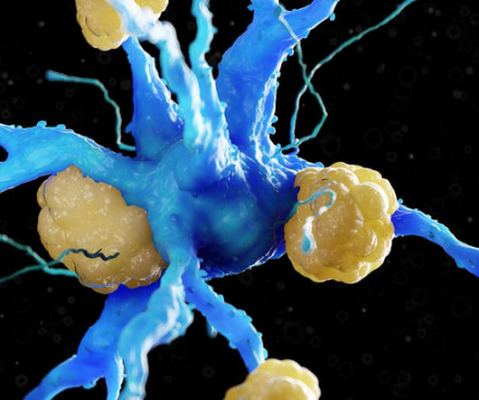
In CAVD, microcrystals of hydroxyapatite (a calcium phosphate mineral) deposit onto the heart valve leaflets and impair cardiacfunction. The disease has a dismal prognosis with most untreated patients dying two years after diagnosis.
Since mitochondrial protein synthesis is critical to its structure, as well as normal cardiacfunction, the authors focused much of their study on how alteration of the mitochondrial protein balance affects heart health.
Cardiac fibrosis is a pathological hallmark of almost all forms of heart disease, characterized by excessive deposition of extracellular matrix (ECM) proteins by activated fibroblasts, leading to cardiomyocyte hypertrophy, arrhythmias, and heart failure.
Currently, no pharmacological interventions have been specifically tailored to treat CH. Furthermore, OTUD7B deficiency exacerbated transverse aortic coarctation surgery‐induced myocardial hypertrophy, abnormal cardiacfunction, and fibrosis.
Abstract Aims Neutrophil activity contributes to adverse cardiac remodelling in experimental acute cardiac injury and is modifiable with pharmacologic agents like colchicine. HFpEF, heart failure with preserved ejection fraction; HFrEF, heart failure with reduced ejection fraction.
Myocardial fibrosis, a common complication of myocardial infarction (MI), is characterized by excessive collagen deposition and can result in impaired cardiacfunction. Remarkably, CD137 knockout mice exhibited improved cardiacfunction and reduced fibrosis compared to wild-type mice at day 28 post-MI.
We investigated the effect of ERR agonist on cardiacfunction in a pressure overload–induced HF model in vivo. Our results provide direct pharmacologic evidence supporting the further development of ERR agonists as novel HF therapeutics.
Ion channels play a crucial role in various aspects of cardiacfunction, such as regulating rhythm and contractility. Cell function is substantially influenced by the concentration of free cytosolic calcium (Ca2+) and the voltage across the plasma membrane.
Furthermore, the differential expression of AMPK/PINK1/Parkin pathway-related proteins in ISO-induced cardiac remodeling was effectively reversed by canagliflozin, suggesting the therapeutic potential of targeting this pathway with the drug. Heart failure has always been a prevalent, disabling, and potentially life-threatening disease.
Serial echocardiography and pressure-volume loops were utilized to assess cardiacfunction and hemodynamics. Serial echocardiography and pressure-volume loops were utilized to assess cardiacfunction and hemodynamics. In contrast, combination therapy resulted in significant improvement.
We found that RUS effectively alleviated myocardial pathological damage, normalized cardiacfunction, and increased survival in septic mice. We found that RUS effectively alleviated myocardial pathological damage, normalized cardiacfunction, and increased survival in septic mice.
PTE administration promoted protective effects in terms of oxidative stress in two experimental models of cardiac diseases: MI and PAH. PTE also improved cardiacfunction in infarcted rats and pulmonary artery flow in animals with PAH. In the PAH model, PTE improved pulmonary artery flow and decreased ROS levels in the lung.
Here, we performed transcriptome analysis of SNPiP-treated mice ventricles to elucidate how SNPiP exerts beneficial effects on cardiacfunction. Here, we performed transcriptome analysis of SNPiP-treated mice ventricles to elucidate how SNPiP exerts beneficial effects on cardiacfunction.
Overexpression of ALKBH5 inhibited H/R-induced cardiomyocyte apoptosis and oxidative stress in vitro, and inhibited I/R-induced collagen deposition, cardiacfunction, and apoptosis in vivo. We observed that ALKBH5 and MG53 were highly expressed in MI. We observed that ALKBH5 and MG53 were highly expressed in MI.
Data express a benefit of SFN treatment on the cardiacfunction of rats with PAH associated with the cellular redox state. Data express a benefit of SFN treatment on the cardiacfunction of rats with PAH associated with the cellular redox state. Catalase and GSH/GSSG ratio were diminished in MCT compared to CTR (P<0.05).
Echocardiography was used to estimate cardiacfunction. Echocardiography was used to estimate cardiacfunction. After 6 weeks, some WT mice (n=24) in CIH group were given sildenafil or saline gavage for another 4 weeks. Blood pressure was regularly measured during the experiment.
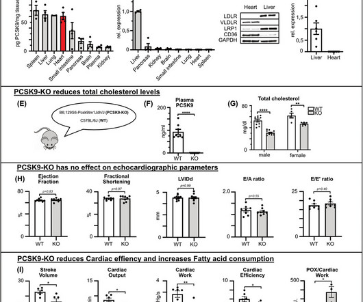
Despite its known effects on cholesterol metabolism, the role of PCSK9 in cardiacfunction, especially post-myocardial infarction (MI), remains unclear. Cardiacfunction was assessed via echocardiography and isolated working heart model experiments. PCSK9 and LDL receptor levels were measured using ELISA and qRT-PCR.
We organize all of the trending information in your field so you don't have to. Join thousands of users and stay up to date on the latest articles your peers are reading.
You know about us, now we want to get to know you!
Let's personalize your content
Let's get even more personalized
We recognize your account from another site in our network, please click 'Send Email' below to continue with verifying your account and setting a password.


Let's personalize your content