ECG Cases 48 – ECG Interpretation in Cardiac Arrest
ECG Cases
FEBRUARY 13, 2024
The post ECG Cases 48 – ECG Interpretation in Cardiac Arrest appeared first on Emergency Medicine Cases.
This site uses cookies to improve your experience. To help us insure we adhere to various privacy regulations, please select your country/region of residence. If you do not select a country, we will assume you are from the United States. Select your Cookie Settings or view our Privacy Policy and Terms of Use.
Cookies and similar technologies are used on this website for proper function of the website, for tracking performance analytics and for marketing purposes. We and some of our third-party providers may use cookie data for various purposes. Please review the cookie settings below and choose your preference.
Used for the proper function of the website
Used for monitoring website traffic and interactions
Cookies and similar technologies are used on this website for proper function of the website, for tracking performance analytics and for marketing purposes. We and some of our third-party providers may use cookie data for various purposes. Please review the cookie settings below and choose your preference.
ECG Cases
FEBRUARY 13, 2024
The post ECG Cases 48 – ECG Interpretation in Cardiac Arrest appeared first on Emergency Medicine Cases.
Dr. Smith's ECG Blog
JULY 9, 2024
Written by Pendell Meyers A man in his 50s was found by his family in cardiac arrest of unknown duration. Despite anticipation by many that the initial post-resuscitation ECG will show an obvious acute infarction — this expected "STEMI picture" is often not seen.
Dr. Smith's ECG Blog
JULY 25, 2024
But cardiac arrest is a period of near zero flow in the coronary arteries and causes SEVERE ischemia. After cardiac arrest, I ALWAYS wait 15 minutes after an ECG like this and record another. See these related cases: Cardiac arrest, defibrillated, diffuse ST depression and ST Elevation in aVR.
Dr. Smith's ECG Blog
SEPTEMBER 20, 2011
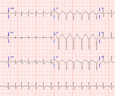
This 80 year old with a history of CABG had a cardiac arrest. This is as clear a STEMI as you can get. Now, it is true that shortly after a non-ACS cardiac arrest, there can be transient diffuse ST depression, but not ST elevation in a coronary distribution, and there should not be a wall motion abnormality.
American College of Cardiology
NOVEMBER 5, 2024
A small proportion of patients with STEMI treated via primary PCI experienced late ventricular tachycardia (VT) or ventricular fibrillation (VF), occurring one or more days following the procedure, but late VT or VF with cardiac arrest occurred rarely, especially among patients with uncomplicated STEMI, according to a study published in JAMA Network (..)
Dr. Smith's ECG Blog
APRIL 24, 2015
ST depression is common BOTH after resuscitation from cardiac arrest and during atrial fib with RVR. Again, it is common to have an ECG that shows apparent subendocardial ischemia after resuscitation from cardiac arrest, after defibrillation, and after cardioversion. The patient was cardioverted. This was done.
Ken Grauer, MD
FEBRUARY 3, 2024
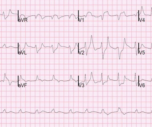
Shortly after arrival in the ED ( E mergency D epartment ) — she suffered a cardiac arrest. BUT — Cardiac catheterization done a little later did not reveal any significant stenosis. Figure-1: The initial ECG in today's case — obtained after successful resuscitation from cardiac arrest. ( No CP ( C hest P ain ).
The British Journal of Cardiology
NOVEMBER 29, 2023
Primary percutaneous coronary intervention (PPCI) remains the gold-standard treatment for ST-elevation myocardial infarction (STEMI). We present the case of a man in his 50s, admitted with cardiac arrest secondary to inferolateral STEMI.
Dr. Smith's ECG Blog
SEPTEMBER 29, 2024
This certainly looks like an anterior STEMI (proximal LAD occlusion), with STE and hyperacute T-waves (HATW) in V2-V6 and I and aVL. How do you explain the anterior STEMI(+)OMI immediately after ROSC evolving into posterior OMI 30 minutes later? This caused a type 2 anterior STEMI.
Dr. Smith's ECG Blog
OCTOBER 28, 2013
The last section is a detailed discussion of the research on aVR in both STEMI and NonSTEMI. The additional ST Elevation in V1 is not usually seen with diffuse subendocardial ischemia, and suggests that something else, like STEMI from LAD occlusion, could be present. Here is an article I wrote: Updates on the ECG in ACS. see below).
Dr. Smith's ECG Blog
OCTOBER 24, 2013
Appreciation of these subtle ECG findings could have helped to avoid a cardiac arrest and its resulting permanent disability 3. Ischemia on the ECG can be very subtle and is easily missed. Accurate interpretation requires a lot of skill, practice, and experience.
Dr. Smith's ECG Blog
OCTOBER 8, 2024
Subtle as a STEMI." (i.e., About 45 minutes after the second EKG, the patient was found in cardiac arrest. Later the next day, she went into cardiac arrest again. By the time I saw the repeat EKG, the patient was already in cardiac arrest. None of the 20 ever evolved to STEMI criteria.
Ken Grauer, MD
JUNE 14, 2024
The ECG in Figure-1 — was obtained from a middle-aged man who presented to the ED ( E mergency D epartment ) in cardiac arrest. Prompt cath is therefore advised if the post-ROSC shows an acute STEMI. To Emphasize: The phenomenon of T-QRS-D is not needed in today's case to recognize the acute STEMI.
Dr. Smith's ECG Blog
JUNE 21, 2023
You can subscribe for news and early access (via participating in our studies) to the Queen of Hearts here: [link] queen-form This EMS ECG was transmitted to the nearby Emergency Department where it was remotely reviewed by a physician, who interpreted it as normal, or at least without any features of ischemia or STEMI.
Dr. Smith's ECG Blog
OCTOBER 4, 2010
Thus, this patient had increased ST elevation (current of injury) superimposed on the ST elevation of LVH and simulating STEMI. The stress of the triathlon cause demand ischemia and ventricular fibrillation. He awoke and did well.
Dr. Smith's ECG Blog
JULY 14, 2023
Two recent interventions have proven in randomized trials to improve neurologic survival in cardiac arrest: 1) the combination of the ResQPod and the ResQPump (suction device for compression-decompression CPR -- Lancet 2011 ) and 2) Dual Sequential defibrillation.
Dr. Smith's ECG Blog
SEPTEMBER 21, 2020
Are Some Cardiologists Really Limited by Strict Adherence to STEMI millimeter criteria? I was texted these ECGs by a recent residency graduate after they had all been recorded, along with the following clinical information: A 50-something with no cardiac history, but with h/o Diabetes, was doing physical work when he collapsed.
Dr. Smith's ECG Blog
AUGUST 19, 2023
This is diagnostic of infero-posterior OMI, but it is falsely negative by STEMI criteria and with falsely negative posterior leads (though they do show mild ST elevation in V4R). They were less likely to have STEMI on ECG, and more likely to be initially diagnosed as non-ACS.
Journal of the American Heart Association
FEBRUARY 19, 2025
BackgroundThere may be variability in willingness to perform percutaneous coronary intervention (PCI) in higherrisk patients who present with STsegmentelevation myocardial infarction (STEMI). There were 178 984 patients from 582 US hospitals presenting with STEMI who were included. versus 7.4%,P<0.001), versus 5.1%,P<0.001)
Dr. Smith's ECG Blog
OCTOBER 17, 2024
Past medical history included RBBB without other cardiac history, but old ECG was not available. The prehospital and ED computer interpretation was inferior STEMI: There’s normal sinus rhythm, first degree AV block and RBBB, normal axis and normal voltages. Vitals were normal except for oxygen saturation of 94%. Vitals were normal.
Dr. Smith's ECG Blog
JUNE 15, 2013
The cath lab was activated for STEMI. Unfortunately, the patient had a cardiac arrest on arrival to the cath lab, before return of the potassium. Hyperkalemia alone can cause inferior-posterior pseudo-STEMI: Notice that in both cases, the ST elevation is downsloping and the T-wave is inverted. The potassium returned at 8.4
Dr. Smith's ECG Blog
DECEMBER 23, 2014
The patient was diagnosed with esophageal reflux and was being discharged by the nurse when he had a cardiac arrest. Anterolateral STEMI. The formula results in 23.43, just above the 23.4 He was defibrillated. Here is his post resuscitation ECG: Now the diagnosis is obvious.
Dr. Smith's ECG Blog
JANUARY 8, 2024
She was diagnosed with a Non-STEMI and kept overnight for a next day angiogram. Medics recorded the above ECG and called a STEMI alert. The patient went into arrest pre-hospital. Her symptoms at the time ECG #1 was obtained were CP and SOB — yet interpretation of this initial tracing prompted a STEMI alert.
Dr. Smith's ECG Blog
NOVEMBER 23, 2022
About an hour later, he was then found on the floor in cardiac arrest in the ED. Sooner identification likely leads to better outcomes, and in this case may have allowed prevention of cardiac arrest and better long-term outcome. His initial troponin T was 15 ng/L (only two hours since pain onset).
Dr. Smith's ECG Blog
FEBRUARY 2, 2017
Discussion See this post: STEMI with Life-Threatening Hypokalemia and Incessant Torsades de Pointes I could find very little literature on the treatment of severe life-threatening hypokalemia. If cardiac arrest from hypokalemia is imminent (i.e., When the ECG shows the effects of hypokalemia, it is particularly dangerous.
Dr. Smith's ECG Blog
NOVEMBER 4, 2022
This has been termed a “STEMI equivalent” and included in STEMI guidelines, suggesting this patient should receive dual anti-platelets, heparin and immediate cath lab activation–or thrombolysis in centres where cath lab is not available. aVR ST segment elevation: acute STEMI or not? aVR ST Segment Elevation: Acute STEMI or Not?
Dr. Smith's ECG Blog
AUGUST 1, 2020
He has done a lot of great work on cardiac arrest, including as co-author of our study on esmolol in refractory cardiac arrest, and much more with Keith Lurie. This was sent by Guillaume Debaty, from the Grenoble Region of France. See his Google Scholar profile here. Several of these are present in this case.
Dr. Smith's ECG Blog
MAY 19, 2020
This is a troponin I level that is almost exclusively seen in STEMI. I suspect this is Type 2 MI due to prolonged severe hypotension from cardiac arrest. So this is either a case of MINOCA, or a case of Type II STEMI. If the arrest was caused by acute MI due to plaque rupture, then the diagnosis is MINOCA.
Dr. Smith's ECG Blog
JUNE 11, 2021
The patient was brought to the ED as a possible Code STEMI and was seen directly by cardiology. Similarly, STEMI guidelines call for urgent angiography for refractory ischemia or electrical/hemodynamic instability, regardless of ECG findings. On arrival, GCS was 13 and the patient complained of ongoing chest pain.
Ken Grauer, MD
OCTOBER 21, 2023
Subendocardial Ischemia from another Cause ( ie, sustained tachyarrhythmia; cardiac arrest; shock or profound hypotension; GI bleeding; anemia; "sick patient" , etc. ). To EMPHASIZE: This pattern of diffuse Subendocardial Ischemia does not suggest acute coronary occlusion ( ie, it is not the pattern of an acute MI ).
Dr. Smith's ECG Blog
DECEMBER 13, 2023
This phenomenon has been associated with cardiac arrest; after cardiac surgery; post-cardioversion following a sustained tachyarrhythmia; with certain types of acute cerebrovascular events ( such as subarachnoid hemorrhage ) — and , in association with large acute MI , which could potentially be the clinical scenario in today's case!
Dr. Smith's ECG Blog
JANUARY 4, 2021
Could this be Septal STEMI (STE in V1 and aVR, with reciprocal ST depression in V4-V6?), In Septal STEMI , transmural ischemia of the septum is recorded by the overlying lead V1 as ST Elevation. Lead III is also on the right and might manifest ST Elevation in Septal STEMI. with ADDED STE in III?
Dr. Smith's ECG Blog
NOVEMBER 30, 2019
If cardiac arrest from hypokalemia is imminent (i.e., to greatly decrease risk (although in STEMI, the optimal level is about 4.0-4.5 As I indicated above, in our cardiac arrest case, after pushing 40 mEq, the K only went up to 4.2 Give an initial infusion of 10 mEq IV over 5 minutes; repeat once if needed.
Dr. Smith's ECG Blog
NOVEMBER 30, 2022
It is apparently fortunate that she had a cardiac arrest; otherwise, her ECG would have been ignored. Then they did an MRI: Patient underwent cardiac MRI on 10/4 that showed mildly reduced BiV systolic function. This has resulted in an under-representation of STEMI MINOCA patients in the literature. From Gue at al.
Dr. Smith's ECG Blog
APRIL 15, 2019
A prehospital ECG was recorded (not shown and not seen by me) which was worrisome for STEMI. A previous ECG from 4 years prior was normal: This looks like an anterior STEMI, but it is complicated by tachycardia (which can greatly elevate ST segments) and by the presentation which is of fever and sepsis.
Dr. Smith's ECG Blog
SEPTEMBER 4, 2016
He had a previous MI with cardiac arrest 2 years prior. Important Learning Point: "STEMI" is defined by millimeter criteria (1 mm in limb leads), which this does not meet. Therefore it is not a STEMI. The ST depression may be the most visibly obvious sign of STEMI. Some are STEMI-equivalents.
Dr. Smith's ECG Blog
OCTOBER 3, 2017
A repeat ECG was done: Obvious anterolateral wall STEMI. This rhythm reportedly produced no palpable pulse, and CPR was continued. 30 seconds later, however, the patient began spontaneously moving and CPR was discontinued. Proximal LAD occlusion with dramatic ST Elevation) The patient was intubated and taken to the cath lab.
Dr. Smith's ECG Blog
FEBRUARY 27, 2023
See many examples of Pseudo STEMI due to hyperkalemia at these two posts: Acute respiratory distress: Correct interpretation of the initial and serial ECG findings, with aggressive management, might have saved his life. No followup EKG was recorded!!
Dr. Smith's ECG Blog
JUNE 27, 2024
A patient had a cardiac arrest with ventricular fibrillation and was successfully defibrillated. COACT: The COACT trial was fatally flawed, and because of it, many cardiologists are convinced that if there are no STEMI criteria, the patient does not need to go to the cath lab. These studies did not address OMI ECG findings!!!
Dr. Smith's ECG Blog
APRIL 8, 2016
Here is his ED ECG: There is obvious infero-posterior STEMI. What are you worried about in addition to his STEMI? Comments: STEMI with hypokalemia, especially with a long QT, puts the patient at very high risk of Torsades or Ventricular fibrillation (see many references, with abstracts, below). There is atrial fibrillation.
Dr. Smith's ECG Blog
NOVEMBER 12, 2020
In a series of 18 patients with COVID and ST elevation, 8 were diagnosed with STEMI, 6 of whom had an angiogram and it showed obstructive coronary disease. 12 All STEMI patients had very high cTn typical of STEMI (cTnT > 1.0
Dr. Smith's ECG Blog
OCTOBER 20, 2013
Note that they finally have laid to rest the new or presumably new LBBB as a criteria for STEMI. Note that they finally have laid to rest the new or presumably new LBBB as a criteria for STEMI. Also note that they allow ST depression c/w posterior MI to be a STEMI equivalent. What is the utility of a head CT in cardiac arrest?
Dr. Smith's ECG Blog
FEBRUARY 22, 2024
A woman in her 50s with dyspnea and bradycardia A patient with cardiac arrest, ROSC, and right bundle branch block (RBBB). HyperKalemia with Cardiac Arrest. Peaked T waves: Hyperacute (STEMI) vs. Early Repolarizaton vs. Hyperkalemia What will you do for this altered and bradycardic patient?
Frontiers in Cardiovascular Medicine
OCTOBER 9, 2024
The included variables were age, pre-hospital cardiac arrest, robust collateral recruitment (Rentrop grade 2 or 3), family history of coronary disease, initial systolic blood pressure, initial heart rate, hypercholesterolemia, culprit vessel, smoking status and TIMI flow pre-PCI. for in-hospital mortality, 0.78 for LVEF < 40%.
Expert insights. Personalized for you.
We have resent the email to
Are you sure you want to cancel your subscriptions?
Let's personalize your content