This site uses cookies to improve your experience. To help us insure we adhere to various privacy regulations, please select your country/region of residence. If you do not select a country, we will assume you are from the United States. Select your Cookie Settings or view our Privacy Policy and Terms of Use.
Cookie Settings
Cookies and similar technologies are used on this website for proper function of the website, for tracking performance analytics and for marketing purposes. We and some of our third-party providers may use cookie data for various purposes. Please review the cookie settings below and choose your preference.
Used for the proper function of the website
Used for monitoring website traffic and interactions
Cookie Settings
Cookies and similar technologies are used on this website for proper function of the website, for tracking performance analytics and for marketing purposes. We and some of our third-party providers may use cookie data for various purposes. Please review the cookie settings below and choose your preference.
Strictly Necessary: Used for the proper function of the website
Performance/Analytics: Used for monitoring website traffic and interactions
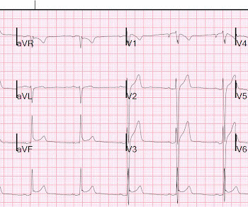
Edited by Bracey, Meyers, Grauer, and Smith A 50-something-year-old female with a history of an unknown personality disorder and alcohol use disorder arrived via EMS following cardiacarrest with return of spontaneous circulation. The described rhythm was an irregular, wide complex rhythm.
The second most common cause of medical cardiac tamponade is acute idiopathic pericarditis. Less common etiologies include uremia, bacterial or tubercular pericarditis, chronic idiopathic pericarditis, hemorrhage, and other causes such as autoimmune diseases, radiation, myxedema, etc.
ECG of pneumopericardium and probable myocardial contusion shows typical pericarditis Male in 30's, 2 days after Motor Vehicle Collsion, complains of Chest Pain and Dyspnea Head On Motor Vehicle Collision. ST depression. Myocardial Contusion?
Further Reading: [link] See these relevant cases: A man in his 50s with acute chest pain and diffuse ST depression "Pericarditis" strikes again Is it important to recognize LVH Pseudo-infarction patterns?
He had multiple cardiacarrests with ROSC regained each time. Dyspnea, Chest pain, Tachypneic, Ill appearing: Bedside Cardiac Echo gives the Diagnosis 31 Year Old Male with RUQ Pain and a History of Pericarditis. Cardiac Ultrasound may be a surprisingly easy way to help make the diagnosis Answer: pulmonary embolism.
This case highlights such a scenario.Case:A 75-year-old female with a history of cardiacarrest 30 years ago presented with shortness of breath and left leg swelling. She experienced massive hemoptysis, leading to respiratory and cardiacarrest, but was resuscitated.
T-wave to ST ratio is greater than 4 in lead V6, making pericarditis unlikely (also there were no symptoms of pericarditis). Sudden cardiacarrest associated with early repolarization. There is ST elevation diffusely: 2 mm in V2, 3.5 mm in V3, 2.5 mm in V4, 1.5 mm in V5, and 1 mm in V6, 1.5 The computerized QTc is 386 ms.
Smith: This bizarre ECG looks like a post cardiacarrest ECG with probable acidosis or hyperkalemia in addition to OMI. Bottom Line: Tests other than cardiac cath may be all that are needed to establish the diagnosis — but, I'd want to see a patient with this ECG as soon as would be possible. What was the pH and K?
They include myocardial ischemia, acute pericarditis, pulmonary embolism, external compression due to mass over the right ventricular outflow tract region, and metabolic disorders like hyper or hypokalemia and hypercalcemia. These are the conditions which have to be considered or excluded as they can sometimes manifest Brugada pattern on ECG.
As always, takotsubo cardiomyopathy and focal pericarditis can mimic OMI, but takotsubo almost never mimics posterior MI, and both are diagnoses of exclusion after a negative cath. About two hours after admission, he suffered a cardiacarrest (whether it was VF/VT or PEA is not available) and expired.
Myocardial rupture is usually preceded by postinfarction regional pericarditis (PIRP). Patients who present with chest pain or cardiacarrest and have an ECG diagnostic of STEMI could have myocardial rupture. In a report of 6 cases at our institution (Hennepin County Medical Center), 2 survived with cardiac surgery.
Differential of peri-infarct pericardial fluid The differential includes 1) pericarditis with effusion or 2) hemopericardium. Differential of peri-infarct pericardial fluid The differential includes 1) pericarditis with effusion or 2) hemopericardium. Dressler's syndrome appears to be quite rare, according to Shahar and Lichstein.
Prominent J waves and ventricular fibrillation caused by myocarditis and pericarditis after BNT162b2 mRNA COVID-19 vaccination. The final letter in the SLOWED mnemonic is " D " for "Dead" ( resulting from VT/VF or asystolic cardiacarrest ). Internat J Arrhyth 2020 Uesako H, Fukikawa H, Hashimoto S, et al.
We organize all of the trending information in your field so you don't have to. Join thousands of users and stay up to date on the latest articles your peers are reading.
You know about us, now we want to get to know you!
Let's personalize your content
Let's get even more personalized
We recognize your account from another site in our network, please click 'Send Email' below to continue with verifying your account and setting a password.


Let's personalize your content