This site uses cookies to improve your experience. To help us insure we adhere to various privacy regulations, please select your country/region of residence. If you do not select a country, we will assume you are from the United States. Select your Cookie Settings or view our Privacy Policy and Terms of Use.
Cookie Settings
Cookies and similar technologies are used on this website for proper function of the website, for tracking performance analytics and for marketing purposes. We and some of our third-party providers may use cookie data for various purposes. Please review the cookie settings below and choose your preference.
Used for the proper function of the website
Used for monitoring website traffic and interactions
Cookie Settings
Cookies and similar technologies are used on this website for proper function of the website, for tracking performance analytics and for marketing purposes. We and some of our third-party providers may use cookie data for various purposes. Please review the cookie settings below and choose your preference.
Strictly Necessary: Used for the proper function of the website
Performance/Analytics: Used for monitoring website traffic and interactions
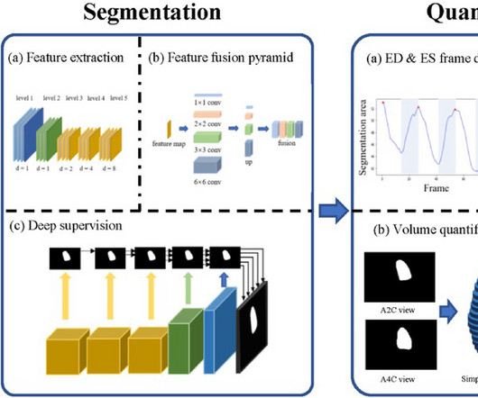
BackgroundPercutaneous extracorporeal membrane oxygenation (ECMO) is administered to pediatric patients with cardiogenic shock or cardiacarrest. Echocardiogram was performed for patients with ECMO, including at pre-ECMO, during cannulation, during ECMO support, during the ECMO wean, and a follow up within 3 months after weaning.
The last section is a detailed discussion of the research on aVR in both STEMI and NonSTEMI. Cardiacarrest can cause diffuse subendocardial ischemia, usually transient (it often resolves as time goes by after ROSC). I repeat that ST elevation in aVR is not diagnostic of left main occlusion. It was stented.
It is apparently fortunate that she had a cardiacarrest; otherwise, her ECG would have been ignored. Here is the cath report: Echocardiogram: There is severe hypokinesis of entire LV apex and apical segment of all the walls. She was defibrillated and resuscitated. I need to innoculate you against the subsequent opinions below.
Arrhythmias: Genetic mutations can also predispose individuals to irregular heart rhythms, such as atrial fibrillation or long QT syndrome, which may increase the risk of stroke or sudden cardiacarrest. Heart imaging, such as echocardiograms or CT scans. Key screenings include: Blood pressure and cholesterol checks.
During the intravenous lacosamide infusion, the patient developed sudden cardiacarrest caused by ventricular arrhythmias necessitating resuscitation. Of note, the patient had a family history of sudden cardiac death. Further research is warranted to investigate the interactions between lacosamide and SCN5A variants.
I remember Allie well from her days in the Research volunteer program at Hennepin. A formal echocardiogram was completed the next day and again showed a normal ejection fraction without any focal wall motion abnormalities to suggest CAD. This was submitted by Alexandra Schick. The article is edited by Smith.
We organize all of the trending information in your field so you don't have to. Join thousands of users and stay up to date on the latest articles your peers are reading.
You know about us, now we want to get to know you!
Let's personalize your content
Let's get even more personalized
We recognize your account from another site in our network, please click 'Send Email' below to continue with verifying your account and setting a password.


Let's personalize your content