This site uses cookies to improve your experience. To help us insure we adhere to various privacy regulations, please select your country/region of residence. If you do not select a country, we will assume you are from the United States. Select your Cookie Settings or view our Privacy Policy and Terms of Use.
Cookie Settings
Cookies and similar technologies are used on this website for proper function of the website, for tracking performance analytics and for marketing purposes. We and some of our third-party providers may use cookie data for various purposes. Please review the cookie settings below and choose your preference.
Used for the proper function of the website
Used for monitoring website traffic and interactions
Cookie Settings
Cookies and similar technologies are used on this website for proper function of the website, for tracking performance analytics and for marketing purposes. We and some of our third-party providers may use cookie data for various purposes. Please review the cookie settings below and choose your preference.
Strictly Necessary: Used for the proper function of the website
Performance/Analytics: Used for monitoring website traffic and interactions
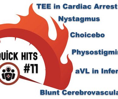
The post EM Quick Hits 11 Blunt Cerebrovascular Injury, Physostigmine, TEE in CardiacArrest, Understanding Nystagmus, Subtle Inferior MI, Choicebo appeared first on Emergency Medicine Cases.
BackgroundPercutaneous extracorporeal membrane oxygenation (ECMO) is administered to pediatric patients with cardiogenic shock or cardiacarrest. Echocardiogram was performed for patients with ECMO, including at pre-ECMO, during cannulation, during ECMO support, during the ECMO wean, and a follow up within 3 months after weaning.
ST depression is common BOTH after resuscitation from cardiacarrest and during atrial fib with RVR. Again, it is common to have an ECG that shows apparent subendocardial ischemia after resuscitation from cardiacarrest, after defibrillation, and after cardioversion. The patient was cardioverted. This was done.
Cardiacarrest can cause diffuse subendocardial ischemia, usually transient (it often resolves as time goes by after ROSC). An echocardiogram on day 3 showed no wall motion abnormality (but of course, these can resolved with reperfusion, and the more time it has to resolve from "stunning", the more likely it is to be resolved).
Echocardiogram An echocardiogram uses sound waves to produce a detailed image of the heart, allowing doctors to see the size of the heart chambers and how well the heart is pumping blood. CardiacArrest or Sudden Death: Cardiomegaly increases the risk of life-threatening arrhythmias, which can cause sudden cardiacarrest.
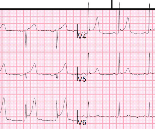
A 35-year-old woman without a history of cardiac disease was seen in the emergency department after an abrupt syncopal episode. Her cardiacechocardiogram and serum electrolytes were normal. She denied the use of any medications, and her family history was unremarkable.
Arrhythmias: Genetic mutations can also predispose individuals to irregular heart rhythms, such as atrial fibrillation or long QT syndrome, which may increase the risk of stroke or sudden cardiacarrest. Heart imaging, such as echocardiograms or CT scans. Key screenings include: Blood pressure and cholesterol checks.
An echocardiogram confirmed aortic stenosis with a large pressure gradient. Now there is much less ST segment deviation, less elevation and less depression. The troponin returned positive, and the maximum troponin was 3.8 The next day, and angiogram showed normal coronary arteries. He awoke and did well.
Two recent interventions have proven in randomized trials to improve neurologic survival in cardiacarrest: 1) the combination of the ResQPod and the ResQPump (suction device for compression-decompression CPR -- Lancet 2011 ) and 2) Dual Sequential defibrillation. Formal Echocardiogram: Normal left ventricular size and wall thickness.
See this post: How a pause can cause cardiacarrest 2. In this specific case, Left Bundle Branch (LBB) area pacing was pursued to achieve cardiac resynchronization. There is ventricular bigeminy with bizarre appearing wide T-waves See even more striking cases of this at the bottom of the post. The plan: 1. J Am Coll Cardiol.
Echocardiogram showed LVEF 66% with normal wall motion and normal diastolic function. However, he did not remember much from the day of the arrest. Lesions less than 70% are generally considered to be non-flow limiting. Two subsequent troponins were down trending.
During the intravenous lacosamide infusion, the patient developed sudden cardiacarrest caused by ventricular arrhythmias necessitating resuscitation. Of note, the patient had a family history of sudden cardiac death.
However, an echocardiogram is a different test, also conducted for heart activity. CardiacarrestCardiacarrest is a medical emergency in which the heart stops pumping blood to the body. Electrocardiogram, echocardiogram, and some other tests are done for patients with cardiacarrest.
Initial evaluation showed elevated cardiac enzymes (CE) and normal eosinophil count. Transthoracic echocardiogram (TTE) showed an ejection fraction (EF) of 40% and a moderate-large pericardial effusion with signs of tamponade. He had a cardiacarrest during the procedure and was placed back on ECMO.
So don't wait for the laboratory K or you might be resuscitating a cardiacarrest ( see the case with ECGs #3 and #4 of this post ). In this study of consecutive patients with LBBB who were hospitalized and had an echocardiogram , 13% had a QRS duration greater than 170 ms, and only 1% had a duration greater than 190 ms.
In any case, the patient needs at a minimum serial ECGs and perhaps a formal echocardiogram. As he was alarmed by it, he went to the patient who now was having recurrent pain, then suddenly went into cardiacarrest (ventricular fibrillation). So, this noise should not be considered TQRSD, and then the formula should not be used.
I think a good start would be a posterior EKG and a high quality contrast echocardiogram read by an expert. Dialysis patients had double the rate of cardiacarrest (11% vs 5%), were less likely to receive reperfusion therapy when eligible (47% vs. 75%), and had an increased odds ratio of death compared to nondialysis patients 1.5 (95%
See this case: what do you think the echocardiogram shows in this case? Thirty-six patients (36%) presented with cardiacarrest, and 78% (28/36) underwent emergent angiography. POCUS showed good LV-function and no pericardial effusion. The patient had mild but diffuse abdominal tenderness.
Formal echocardiogram showed normal EF, no wall motion abnormalities, no pericardial effusion. The patient proceeded to cath where all coronaries were described as normal with no evidence of any CAD, spasm, or any other abnormality. No more troponins were done. He was found to be influenza positive. 1849 after cath: Brugada pattern is gone!
hours ECG: Not much change hs troponin I peaks at 500 ng/L 8 hours Next morning Urine drug screen: Amphetamine, Methamphetamine, Fentanyl, Fentanyl metabolite Formal Bubble Contrast Echocardiogram: Indications for Study: Silent Ischemia. SUMMARY Normal left ventricular cavity size. Normal estimated left ventricular ejection fraction.
Tortuous LAD consistent with hypertensive cardiac disease and luminal irregularities, but free of stenosis 3. Echocardiogram findings (pre-procedure) 1. Had such been the case, this patient would likely have been a prehospital cardiacarrest, or have been in profound cardiogenic shock at the time of ED arrival.
A formal echocardiogram was completed the next day and again showed a normal ejection fraction without any focal wall motion abnormalities to suggest CAD. Cardiology was consulted and they agreed that the EKG had an atypical morphology for STEMI and did not activate the cath lab.
An echocardiogram was done. These include ( among others ) — acute febrile illness — variations in autonomic tone — hypothermia — ischemia-infarction — malignant arrhythmias — cardiacarrest — and especially Hyperkalemia. Is there also Brugada? Here is the result: The estimated left ventricular ejection fraction is 50 %.
It is apparently fortunate that she had a cardiacarrest; otherwise, her ECG would have been ignored. Here is the cath report: Echocardiogram: There is severe hypokinesis of entire LV apex and apical segment of all the walls. She was defibrillated and resuscitated. I need to innoculate you against the subsequent opinions below.
I have ordered an echocardiogram which will be done today, after that patient can be discharged to home with follow-up in 2 to 3 months." The echo was normal. Learning points 1. In this regular wide complex tachycardia , since the rhythm converted w adenosine, it is almost certainly SVT w aberrancy, which can be either: A.
Patients who present with chest pain or cardiacarrest and have an ECG diagnostic of STEMI could have myocardial rupture. In a report of 6 cases at our institution (Hennepin County Medical Center), 2 survived with cardiac surgery. In contrast to re-occlusion of the infarct-related artery, this reversal should be gradual.
Smith comment: The patient was lucky to have a cardiacarrest. By undergoing an arrest, providers became aware of his OMI which had not been noticed on his diagnostic ECG, and he thus has a chance at some myocardial salvage. Had he not had one, he would have sat in the waiting room until his entire myocardium at risk infarcted.
In this case, a patient experienced sudden cardiacarrest during sexual activity, which has not previously been reported.Case presentationSix years ago, a 37-year-old man was admitted with sudden cardiacarrest during sexual intercourse. No previous history of hypertension or diabetes.
We organize all of the trending information in your field so you don't have to. Join thousands of users and stay up to date on the latest articles your peers are reading.
You know about us, now we want to get to know you!
Let's personalize your content
Let's get even more personalized
We recognize your account from another site in our network, please click 'Send Email' below to continue with verifying your account and setting a password.


Let's personalize your content