This site uses cookies to improve your experience. To help us insure we adhere to various privacy regulations, please select your country/region of residence. If you do not select a country, we will assume you are from the United States. Select your Cookie Settings or view our Privacy Policy and Terms of Use.
Cookie Settings
Cookies and similar technologies are used on this website for proper function of the website, for tracking performance analytics and for marketing purposes. We and some of our third-party providers may use cookie data for various purposes. Please review the cookie settings below and choose your preference.
Used for the proper function of the website
Used for monitoring website traffic and interactions
Cookie Settings
Cookies and similar technologies are used on this website for proper function of the website, for tracking performance analytics and for marketing purposes. We and some of our third-party providers may use cookie data for various purposes. Please review the cookie settings below and choose your preference.
Strictly Necessary: Used for the proper function of the website
Performance/Analytics: Used for monitoring website traffic and interactions
A 60-something woman presented after a witnessed cardiacarrest. This is commonly found after epinephrine for cardiacarrest, but could have been pre-existing and a possible contributing factor to cardiacarrest. Final Diagnosis: CardiacArrest due to Torsades from long QT of unknown etiology.
His response: “subendocardial ischemia. Smith : It should be noted that, in subendocardial ischemia, in contrast to OMI, absence of wall motion abnormality is common. With the history of Afib, CTA abdomen was ordered to r/o mesenteric ischemia vs ischemic colitis vs small bowel obstruction. Anything more on history?
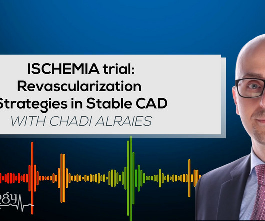
In the ISCHEMIA (International Study of Comparative Health Effectiveness with Medical and Invasive Approaches) trial, researchers examined the risk of ischemic events in patients with stable coronaryarterydisease. years, with 57.1% occurring within 30 days after CABG. Original article: Redfors B et al.
It is apparently fortunate that she had a cardiacarrest; otherwise, her ECG would have been ignored. Hospital Course The patient was taken emergently to the cath lab which did not reveal any significant coronaryarterydisease, but she was noted to have reduced EF consistent with Takotsubo cardiomyopathy.
Heart Valve Disease If one or more heart valves are not functioning correctly, it can cause blood to flow backward, putting extra pressure on the heart, which may cause it to expand to compensate for the inefficiency. This may result in ischemia (lack of oxygen to the heart muscle), causing parts of the heart to weaken and enlarge.
The first task when assessing a wide complex QRS for ischemia is to identify the end of the QRS. The ST segment changes are compatible with severe subendocardial ischemia which can be caused by type I MI from ACS or potentially from type II MI (non-obstructive coronaryarterydisease with supply/demand mismatch).
Atherosclerotic cardiovascular disease (ASCVD), caused by plaque buildup in arterial walls, is one of the leading causes of disability and death worldwide.1,2 1,2 ASCVD causes or contributes to conditions that include coronaryarterydisease (CAD), cerebrovascular disease, and peripheral vascular disease (inclusive of aortic aneurysm).3
CardiacarrestCardiacarrest is a medical emergency in which the heart stops pumping blood to the body. Electrocardiogram, echocardiogram, and some other tests are done for patients with cardiacarrest. ECG machine can also help diagnose coronaryarterydisease.
There is no definite evidence of acute ischemia. (ie, Simply stated — t he patient was having recurrent PMVT without Q Tc prolongation, and without evidence of ongoing transmural ischemia. ( If there had been ECG findings indicating reocclusion of the artery — an angiogram would have been warranted ).
The highly impactful International Study of Comparative Health Effectiveness With Medical and Invasive Approaches ( ISCHEMIA ) trial investigated the effectiveness of invasive (INV) versus conservative (CON) strategies for managing stable coronaryarterydisease. Original article: Gaudino M et al. EuroIntervention.
We evaluated the primary outcome (cardiovascular death, myocardial infarction, or hospitalization for unstable angina, heart failure, or resuscitated cardiacarrest) and other end points, by sex, in 1168 (22.6%) women and 4011 (77.4%) men. Women had less coronaryarterydisease: multivessel in 60.0%
We organize all of the trending information in your field so you don't have to. Join thousands of users and stay up to date on the latest articles your peers are reading.
You know about us, now we want to get to know you!
Let's personalize your content
Let's get even more personalized
We recognize your account from another site in our network, please click 'Send Email' below to continue with verifying your account and setting a password.


Let's personalize your content