This site uses cookies to improve your experience. To help us insure we adhere to various privacy regulations, please select your country/region of residence. If you do not select a country, we will assume you are from the United States. Select your Cookie Settings or view our Privacy Policy and Terms of Use.
Cookie Settings
Cookies and similar technologies are used on this website for proper function of the website, for tracking performance analytics and for marketing purposes. We and some of our third-party providers may use cookie data for various purposes. Please review the cookie settings below and choose your preference.
Used for the proper function of the website
Used for monitoring website traffic and interactions
Cookie Settings
Cookies and similar technologies are used on this website for proper function of the website, for tracking performance analytics and for marketing purposes. We and some of our third-party providers may use cookie data for various purposes. Please review the cookie settings below and choose your preference.
Strictly Necessary: Used for the proper function of the website
Performance/Analytics: Used for monitoring website traffic and interactions
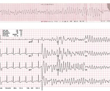
See this post: How a pause can cause cardiacarrest 2. Finally, do a coronaryangiogram Possible alternative to pacing is to give a beta-1 agonist to increase heart rate. In this specific case, Left Bundle Branch (LBB) area pacing was pursued to achieve cardiac resynchronization. Place temporary pacemaker 3.
Initial evaluation showed elevated cardiac enzymes (CE) and normal eosinophil count. Transthoracic echocardiogram (TTE) showed an ejection fraction (EF) of 40% and a moderate-large pericardial effusion with signs of tamponade. A repeat coronaryangiogram was unremarkable. Electrocardiogram (EKG) was unremarkable.
It’s judicious, then, to arrange for coronaryangiogram. Coronary occlusion, however, might be present concurrently with subendocardial ischemia on the time-zero ECG, or evolve into such. Proximal LAD disease with/without a) and b) It seemed quite apparent that this was an Acute Coronary Syndrome. CoronaryAngiogram 1.
See this case: what do you think the echocardiogram shows in this case? We investigated the incidence of an acutely occluded coronary in patients presenting with STE-aVR with multi-lead ST depression. All electrocardiograms (ECGs) and coronaryangiograms were blindly analyzed by experienced cardiologists.
During the intravenous lacosamide infusion, the patient developed sudden cardiacarrest caused by ventricular arrhythmias necessitating resuscitation. Of note, the patient had a family history of sudden cardiac death.
We organize all of the trending information in your field so you don't have to. Join thousands of users and stay up to date on the latest articles your peers are reading.
You know about us, now we want to get to know you!
Let's personalize your content
Let's get even more personalized
We recognize your account from another site in our network, please click 'Send Email' below to continue with verifying your account and setting a password.


Let's personalize your content