This site uses cookies to improve your experience. To help us insure we adhere to various privacy regulations, please select your country/region of residence. If you do not select a country, we will assume you are from the United States. Select your Cookie Settings or view our Privacy Policy and Terms of Use.
Cookie Settings
Cookies and similar technologies are used on this website for proper function of the website, for tracking performance analytics and for marketing purposes. We and some of our third-party providers may use cookie data for various purposes. Please review the cookie settings below and choose your preference.
Used for the proper function of the website
Used for monitoring website traffic and interactions
Cookie Settings
Cookies and similar technologies are used on this website for proper function of the website, for tracking performance analytics and for marketing purposes. We and some of our third-party providers may use cookie data for various purposes. Please review the cookie settings below and choose your preference.
Strictly Necessary: Used for the proper function of the website
Performance/Analytics: Used for monitoring website traffic and interactions
Two recent interventions have proven in randomized trials to improve neurologic survival in cardiacarrest: 1) the combination of the ResQPod and the ResQPump (suction device for compression-decompression CPR -- Lancet 2011 ) and 2) Dual Sequential defibrillation. Formal Echocardiogram: Normal left ventricular size and wall thickness.
Case presentation:A 64-year-old man presented with one day of chestpain. Initial evaluation showed elevated cardiac enzymes (CE) and normal eosinophil count. Transthoracic echocardiogram (TTE) showed an ejection fraction (EF) of 40% and a moderate-large pericardial effusion with signs of tamponade.
This patient, who is a mid 60s female with a history of hypertension, hyperlipidemia and GERD, called 911 because of chestpain. A mid 60s woman with history of hypertension, hyperlipidemia, and GERD called 911 for chestpain. It is also NOT the clinical scenario of takotsubo (a week of intermittent chestpain).
The best course is to wait until the anatomy is defined by angio, then if proceeding to PCI, add Cangrelor (an IV P2Y12 inhibitor) I sent the ECG and clinical information of a 90-year old with chestpain to Dr. McLaren. See this case: what do you think the echocardiogram shows in this case?
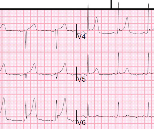
But the symptoms returned with similar pattern – provoked by exertion, and alleviated with rest; except that on each occasion the chestpain was a little more intense, and the needed recovery period was longer in duration. Tortuous LAD consistent with hypertensive cardiac disease and luminal irregularities, but free of stenosis 3.
The pneumothorax was expanded with a chest tube At 17 hours, another ECG was recorded: It is now much less dramatic and has the morphology of Type 2 Brugada The hs troponin I peaked at 6500 ng/L -- this strongly suggests myocardial contusion. An echocardiogram was done. Is there also Brugada? Right ventricular prominence.
Echocardiogram showed LVEF 66% with normal wall motion and normal diastolic function. However, he did not remember much from the day of the arrest. He did not remember whether he had experienced any chestpain. Two subsequent troponins were down trending.
Chest X-Ray A chest X-ray is often the first imaging test conducted, as it can reveal whether the heart is enlarged and by how much. Echocardiogram An echocardiogram uses sound waves to produce a detailed image of the heart, allowing doctors to see the size of the heart chambers and how well the heart is pumping blood.
He complained of severe chestpain and was extremely agitated, so much so that he was throwing chairs in triage. Some time later, reperfusion T-waves developed (analogous to Wellens' waves): Case 3 Here is a case of a 30-something otherwise healthy male with chestpain: There is neither an S-wave nor J-wave in lead V3.
However, an echocardiogram is a different test, also conducted for heart activity. If you experience any symptoms, such as chestpain, dizziness, unusual tiredness or fatigue, shortness of breath, or irregular heartbeat, your doctor would want you to go for an ECG test to find out the underlying cause.
Written by Pendell Meyers A man in his 70s with no cardiac history presented with acute weakness, syncope, and fever. He denied chestpain or shortness of breath. In the clinical context of weakness and fever, without chestpain or shortness of breath, the likelihood of Brugada pattern is obviously much higher.
No chestpain. I think a good start would be a posterior EKG and a high quality contrast echocardiogram read by an expert. His inpatient clinicians did not think that an urgent angiogram was warranted given that he was chestpain free, his EKG appeared nondiagnostic, and serial troponins were not elevating beyond 2 ug/L.
A formal echocardiogram was completed the next day and again showed a normal ejection fraction without any focal wall motion abnormalities to suggest CAD. It was from a patient with chestpain: Note the obvious Brugada pattern. The Troponin I was cycled over time and was 0.353 followed by 0.296. This patient ruled out for MI.
Patients who present with chestpain or cardiacarrest and have an ECG diagnostic of STEMI could have myocardial rupture. In a report of 6 cases at our institution (Hennepin County Medical Center), 2 survived with cardiac surgery. Obviously, administration of heparin and/or lytics is hazardous.
Written by Pendell Meyers A man in his 60s presented with acute chestpain. Smith comment: The patient was lucky to have a cardiacarrest. By undergoing an arrest, providers became aware of his OMI which had not been noticed on his diagnostic ECG, and he thus has a chance at some myocardial salvage.
We organize all of the trending information in your field so you don't have to. Join thousands of users and stay up to date on the latest articles your peers are reading.
You know about us, now we want to get to know you!
Let's personalize your content
Let's get even more personalized
We recognize your account from another site in our network, please click 'Send Email' below to continue with verifying your account and setting a password.


Let's personalize your content