This site uses cookies to improve your experience. To help us insure we adhere to various privacy regulations, please select your country/region of residence. If you do not select a country, we will assume you are from the United States. Select your Cookie Settings or view our Privacy Policy and Terms of Use.
Cookie Settings
Cookies and similar technologies are used on this website for proper function of the website, for tracking performance analytics and for marketing purposes. We and some of our third-party providers may use cookie data for various purposes. Please review the cookie settings below and choose your preference.
Used for the proper function of the website
Used for monitoring website traffic and interactions
Cookie Settings
Cookies and similar technologies are used on this website for proper function of the website, for tracking performance analytics and for marketing purposes. We and some of our third-party providers may use cookie data for various purposes. Please review the cookie settings below and choose your preference.
Strictly Necessary: Used for the proper function of the website
Performance/Analytics: Used for monitoring website traffic and interactions
Sent by Magnus Nossen MD, written by Pendell Meyers A man in his 50s, previously healthy, developed acute chestpain. The primary care physician there evaluated this patient and deemed the chestpain to be due to gastrointestinal causes. The ECG was also interpreted as normal by the primary care physician.
The chestpain quickly subsided. Cardiacarrest was called and advanced life support was undertaken for this patient. The patient was given chest compressions while waiting for the cardiacarrest team to arrive. After about 90 seconds of chest compressions she awoke. Calcium level was normal.
But cardiacarrest is a period of near zero flow in the coronary arteries and causes SEVERE ischemia. After cardiacarrest, I ALWAYS wait 15 minutes after an ECG like this and record another. Just as important is pretest probability: did the patient report chestpain prior to collapse?
Written by Jesse McLaren Two patients in their 70s presented to the ED with chestpain and RBBB. Patient 1 : a 75 year old called paramedics with one day of left shoulder pain which migrated to the central chest, which was worse with deep breaths. Do either, both, or neither have occlusion MI? Vitals were normal.
Let me tell you about her hospitalization, discharged 1 day prior, but it was at another hospital (I wish I had the ECG from that hospitalization): The patient is 40 years old and presented to another hospital with chestpain and SOB. She had been sitting doing work when she experienced "waves of chest tightness". Sats were 88%.
Case submitted by Rachel Plate MD, written by Pendell Meyers A man in his 70s presented with chestpain which had started acutely at rest and has lasted for 2 hours. The pain was still ongoing at arrival. His initial troponin T was 15 ng/L (only two hours since pain onset). He stated it was similar to prior heart attacks.
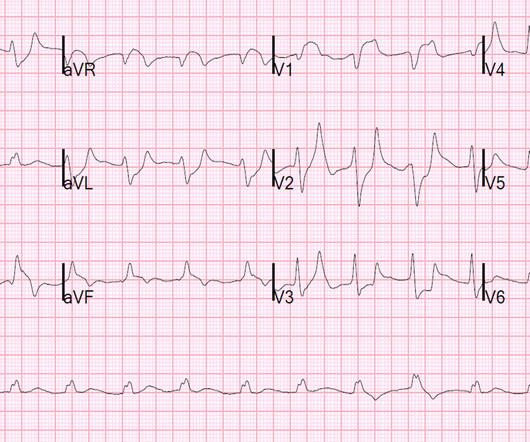
There is no way to tell the difference between GI etiology of chestpain and MI. Such T-waves are almost always reciprocal to ischemia in the region of aVL (although aVL looks n ormal here) , and in a patient with chestpain are nearly diagnostic of ischemia. Could this have been avoided? Lesson : 1.
This is another case written by Pendell Meyers (who is helping to edit the blog and has many great recent posts) Case A 45 year old man was driving to work when he experienced acute onset sharp left sided chestpain with paresthesias of the left arm. A repeat ECG was recorded with pain 2/10: Not much change.
The patient presented due to chestpain that was typical in nature, retrosternal and radiating to the left arm and neck. He denied any exertional chestpain. It is unclear if the patient was pain free at this time. He has a medical hx notable for hypertension, hyperlipidemia and previous tobacco use disorder.
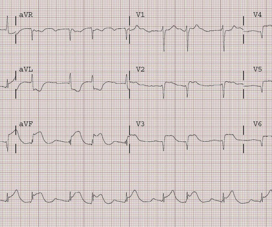
This 80 year old with a history of CABG had a cardiacarrest. He did not state he had chestpain, but, then again, he couldn't remember anything. He was resuscitated after fairly prolonged down time, but regained consciousness, though he was confused. There is concordant ST elevation in all inferior leads.
The patient was diagnosed with esophageal reflux and was being discharged by the nurse when he had a cardiacarrest. Formula : There is not enough ST elevation in V2-V4 to be applying the LAD/early repol formula, but if it is applied, one gets 1.5 The formula results in 23.43, just above the 23.4 He was defibrillated.
A small proportion of patients with STEMI treated via primary PCI experienced late ventricular tachycardia (VT) or ventricular fibrillation (VF), occurring one or more days following the procedure, but late VT or VF with cardiacarrest occurred rarely, especially among patients with uncomplicated STEMI, according to a study published in JAMA Network (..)
Patients may feel a fluttering in the chest, chestpain, shortness of breath and dizziness or lightheadedness as a result. If VT is not treated, it can lead to cardiacarrest, which is when the heart stops beating. In fact, VT is the most common cause of sudden cardiacarrest.
Two recent interventions have proven in randomized trials to improve neurologic survival in cardiacarrest: 1) the combination of the ResQPod and the ResQPump (suction device for compression-decompression CPR -- Lancet 2011 ) and 2) Dual Sequential defibrillation.
Because the patient had no chestpain or shortness of breath, they were initially diagnosed as gastroenteritis. But because the patient had no chestpain or shortness of breath, it was not deemed to be from ACS. Potassium was normal. Cardiology did not think it was "STEMI", but repeated the troponin. Take home 1.
A 60-something woman presented after a witnessed cardiacarrest. This is commonly found after epinephrine for cardiacarrest, but could have been pre-existing and a possible contributing factor to cardiacarrest. A recent similar case: A 40-something with chestpain. Is this inferior MI?
Submitted and written by Quinton Nannet, MD, peer reviewed by Meyers, Grauer, Smith A woman in her 70s recently diagnosed with COVID was brought in by EMS after she experienced acute onset sharp midsternal chestpain without radiation or dyspnea. She felt nauseous and lightheaded with no neurologic deficits.
Shortly after arrival in the ED ( E mergency D epartment ) — she suffered a cardiacarrest. BUT — Cardiac catheterization done a little later did not reveal any significant stenosis. Figure-1: The initial ECG in today's case — obtained after successful resuscitation from cardiacarrest. ( No CP ( C hest P ain ).
A 56 yo f with h/o HTN and hypercholesterolemia called EMS from home after onset of L chestpain radiating to the left arm. Before EMS arrived, she had "seizure activity" and became unresponsive. She was defibrillated successfully from ventricular fibrillation and developed a perfusing rhythm. She was intubated.
BACKGROUND:There is no specific treatment for sudden cardiacarrest (SCA) manifesting as pulseless electric activity (PEA) and survival rates are low; unlike ventricular fibrillation (VF), which is treatable by defibrillation. Circulation: Arrhythmia and Electrophysiology, Ahead of Print.
The ECG in Figure-1 — was obtained from a middle-aged man who presented to the ED ( E mergency D epartment ) in cardiacarrest. C ASE C onclusion: As noted above — the middle-aged man in today's case presented to the ED in cardiacarrest. In view of this history — How would YOU interpret the ECG in Figure-1 ?
Subendocardial Ischemia from another Cause ( ie, sustained tachyarrhythmia; cardiacarrest; shock or profound hypotension; GI bleeding; anemia; "sick patient" , etc. ). Having looked for negative U waves in patients with chestpain over a period of decades — I'll emphasize that this is not a common finding.
Jesse McLaren (@ECGcases), of Emergency Medicine Cases Reviewed by Pendell Meyers and Steve Smith An 85yo with a history of hypertension developed chestpain and collapsed, and had bystander CPR. On arrival, GCS was 13 and the patient complained of ongoing chestpain. Vitals were HR 58 BP 167/70 R20 sat 96%.
There was no chestpain. Unfortunately, the patient had a cardiacarrest on arrival to the cath lab, before return of the potassium. A patient who does not present with chestpain should be particularly scrutinized for other causes of the ECG abnormalities. The computer read inferior MI.
While many arrhythmias are harmless, some can be life-threatening and increase your risk of stroke, heart failure, and sudden cardiacarrest. This can lead to chestpain (angina) and increase your risk of heart attack or stroke, especially if you already have underlying heart disease.
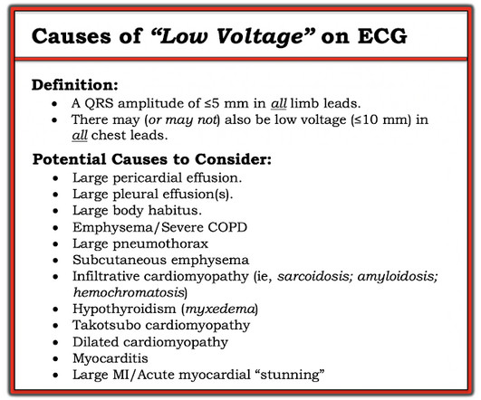
This middle aged patient presented with chestpain: What do you think? There is VERY low voltage. There is some ST Elevation, but it is MINIMAL. But look how small the QRS is!! Let's stretch out the QRS vertically so it is not so tiny: On upper left is the original.
Case presentation:A 64-year-old man presented with one day of chestpain. Initial evaluation showed elevated cardiac enzymes (CE) and normal eosinophil count. He had a cardiacarrest during the procedure and was placed back on ECMO. Circulation, Volume 150, Issue Suppl_1 , Page A4135360-A4135360, November 12, 2024.
He has done a lot of great work on cardiacarrest, including as co-author of our study on esmolol in refractory cardiacarrest, and much more with Keith Lurie. A 40-something woman was in a remote alpine location when she complained of crushing chestpain. See his Google Scholar profile here.
No chestpain. Figure-1: The initial ECG in today's case — obtained from an 86-year old man with presyncope, but no chestpain. ( The 12-lead ECG and long lead II rhythm strip in Figure-1 was obtained from an 86-year old man — who presented to the ED ( E mergency D epartment ) with presyncope. What is the rhythm ?
This patient, who is a mid 60s female with a history of hypertension, hyperlipidemia and GERD, called 911 because of chestpain. A mid 60s woman with history of hypertension, hyperlipidemia, and GERD called 911 for chestpain. It is also NOT the clinical scenario of takotsubo (a week of intermittent chestpain).
When discussing heart health, heart attacks and cardiacarrest are two terms that are often mistaken for one another. Understanding the difference between heart attack and cardiacarrest can help in recognizing symptoms, seeking prompt medical care, and even saving lives. What is CardiacArrest?
The best course is to wait until the anatomy is defined by angio, then if proceeding to PCI, add Cangrelor (an IV P2Y12 inhibitor) I sent the ECG and clinical information of a 90-year old with chestpain to Dr. McLaren. Thirty-six patients (36%) presented with cardiacarrest, and 78% (28/36) underwent emergent angiography.
Case A 47 year old male called 911 for severe chestpain. He had a previous MI with cardiacarrest 2 years prior. A woman in her 60s with no prior history of CAD presented with 3 hours of sharp, centrally located chestpain with radiation to the anterior neck, with associated nausea.
FYI : 52 ng/L is the threshold for "rule in" by European studies as it has a high positive predictive value in the setting of chestpain. Case continued The patient was placed on a nitroglycerin drip and chestpain gradually resolved. Top right is colored iodine overlay; Blue areas of myocardium are ischemia.
If a patient presents with chestpain and a normal heart rate, or with shockable cardiacarrest, then ischemic appearing ST elevation is STEMI until proven otherwise. It is important to remember that not every acute MI with ST elevation is the result of acute coronary occlusio n. Clinical Context is everything !
And she does not know that this is an overdose; she thinks it is a patient with chestpain!! This meets the Smith Modified Sgarbossa criteria, but the situation is wrong for diagnosing OMI!! By the way, the PM Cardio Bot Queen of Hearts says this is Not OMI with High Confidence. 3 hours later, this was recorded at a K of 2.8
Given the history of dyspnea on exertion over a several week period ( but no mention of chestpain ) — and — the finding of deep, symmetric T wave inversion in the anterior leads ( as per Pearl #2 ) — it is possible that the onset of her symptoms is the result of a "Silent MI" ( See ECG Blog #228 for more on "Silent" MI ). . =
This is often quite challenging to recognize — but the finding of negative U waves in a patient with chestpain is highly suggestive of ischemia ! Anterior leads, such as V2 and V3 — as well as lead II — often show U waves best, though they may appear in any lead. N OTE # 2 — On rare occasions, the U wave may be negative.
But the symptoms returned with similar pattern – provoked by exertion, and alleviated with rest; except that on each occasion the chestpain was a little more intense, and the needed recovery period was longer in duration. Then, she attempted to reengage the activities at hand, and initially tolerated this well.
ECG of pneumopericardium and probable myocardial contusion shows typical pericarditis Male in 30's, 2 days after Motor Vehicle Collsion, complains of ChestPain and Dyspnea Head On Motor Vehicle Collision. Gunshot wound to the chest with ST Elevation Would your radiologist make this diagnosis, or should you record an ECG in trauma?
LEARNING Point: Maximal ST depression in leads V2-thru-V4 ( especially when the ST-T waves are shaped as they are in ECG #1 ) in a patient with new chestpain ( or sudden cardiacarrest, as in today’s case ) — is diagnostic of acute Posterior OMI until proven otherwise!
Photo by Cedars-Sinai milla1cf Fri, 03/01/2024 - 08:25 March 1, 2024 — Two new studies by Cedars-Sinai investigators support using artificial intelligence (AI) to predict sudden cardiacarrest-a health emergency that in 90% of cases leads to death within minutes.
However, he did not remember much from the day of the arrest. He did not remember whether he had experienced any chestpain. Within a few days, the patient was extubated and was neurologically intact. At his family's request, he was transferred to a hospital closer to his home to continue care. He was admitted to cardiology.
Case submitted by Magnus Nossen MD from Norway, written by Pendell Meyers A man in his 50s with no pertinent medical history suffered a witnessed cardiacarrest. 12 minutes later, the patient went back into VFib arrest and underwent another 15 minutes of resuscitation followed by successful defibrillation and sustained ROSC.
We organize all of the trending information in your field so you don't have to. Join thousands of users and stay up to date on the latest articles your peers are reading.
You know about us, now we want to get to know you!
Let's personalize your content
Let's get even more personalized
We recognize your account from another site in our network, please click 'Send Email' below to continue with verifying your account and setting a password.


Let's personalize your content