This site uses cookies to improve your experience. To help us insure we adhere to various privacy regulations, please select your country/region of residence. If you do not select a country, we will assume you are from the United States. Select your Cookie Settings or view our Privacy Policy and Terms of Use.
Cookie Settings
Cookies and similar technologies are used on this website for proper function of the website, for tracking performance analytics and for marketing purposes. We and some of our third-party providers may use cookie data for various purposes. Please review the cookie settings below and choose your preference.
Used for the proper function of the website
Used for monitoring website traffic and interactions
Cookie Settings
Cookies and similar technologies are used on this website for proper function of the website, for tracking performance analytics and for marketing purposes. We and some of our third-party providers may use cookie data for various purposes. Please review the cookie settings below and choose your preference.
Strictly Necessary: Used for the proper function of the website
Performance/Analytics: Used for monitoring website traffic and interactions
Cardiacarrest was called and advanced life support was undertaken for this patient. The patient was given chest compressions while waiting for the cardiacarrest team to arrive. She spontaneously converted (Defibrillation was not performed). The morning before the cardiacarrest potassium was 4,3.mmol,
He was defibrillated into VT. He then underwent dual sequential defibrillation into asystole. But cardiacarrest is a period of near zero flow in the coronary arteries and causes SEVERE ischemia. After cardiacarrest, I ALWAYS wait 15 minutes after an ECG like this and record another. They started CPR.
He was defibrillated, but they also noticed that he was being internally defibrillated and then found that he had an implantable ICD. He was unidentified and there were no records available After 7 shocks, he was successfully defibrillated and brought to the ED. I wrote the following note in the chart: "V Fib arrest, has ICD.
While on telemetry monitoring he suffered cardiacarrest and was resuscitated. What ECG finding may have contributed to (or precipitated) the cardiacarrest? After resuscitation and defibrillation , there were no more episodes of TdP. Below is the patient’s 12 lead ECG following defibrillation.
Hypertrophic cardiomyopathy (HCM) predisposes patients to cardiacarrest (CA). While established major risk factors may warrant the need for primary prevention by implantable cardioverter-defibrillator (ICD), it is unknown if specific electrocardiographic predictors increase the risk of CA.
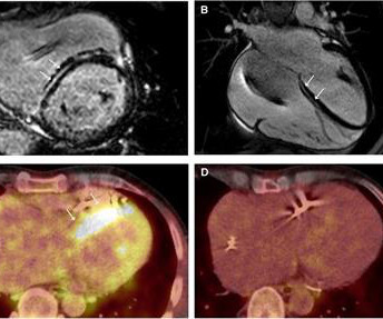
Cardiac sarcoidosis (CS), a rare condition characterized by non-caseating granulomas, can manifest with symptoms such as atrioventricular block and ventricular tachycardia (VT), as well as mimic inherited cardiomyopathies. An endomyocardial biopsy (EMB) confirmed the diagnosis of cardiac sarcoidosis.
And of course Ken's comments at the bottom) An elderly obese woman with cardiomyopathy, Left bundle branch block, and chronic hypercapnea presented hypoxic with altered mental status. Bedside cardiac ultrasound showed moderately decreased LV function. See this post: How a pause can cause cardiacarrest 2. The plan: 1.
She was never seen to be in ventricular fibrillation and was never defibrillated. What is the utility of a head CT in cardiacarrest? We found intracranial hemorrhage in 2% of non-traumatic cardiacarrest patients, and in 4 others the presence of cerebral edema changed management. BP gradually rose.
CardiomyopathyCardiomyopathy is a condition that affects the heart muscle, causing it to become enlarged, thick, or rigid. Excessive Alcohol or Drug Use Long-term abuse of alcohol or certain drugs can weaken the heart muscle, resulting in cardiomyopathy and eventually cardiomegaly.
Although one may have all kinds of ischemic findings as a result of cardiacarrest (rather than cause of cardiacarrest), this degree of ST elevation and HATW is all but diagnostic of acute proximal LAD occlusion. This prompted cath lab activation. On arrival to the ED, this ECG was recorded: What do you think?
Edited by Bracey, Meyers, Grauer, and Smith A 50-something-year-old female with a history of an unknown personality disorder and alcohol use disorder arrived via EMS following cardiacarrest with return of spontaneous circulation. She was successfully revived after several rounds of ACLS including defibrillation and amiodarone.
Video-based AI A profound learning approach is created with a video-based neural system that utilizes a current database of video formats to determine cardiac issues. The deep learning algorithm helps segment the left ventricle predicting cardiomyopathy and ejection fraction. AI recognizing cardiacarrests in emergency calls.
She was defibrillated and resuscitated. It is apparently fortunate that she had a cardiacarrest; otherwise, her ECG would have been ignored. by making it clear to everyone that this is NOT an EKG that one sees with takotsubo cardiomyopathy. Smith: this ECG and clinical presentation is diagnostic of LAD Occlusion.
BACKGROUND:Sudden cardiac death is the most common cause of death in childhood hypertrophic cardiomyopathy (HCM). Recently, 2 risk scores have been developed to estimate the 5-year risk of sudden cardiac death. Circulation: Arrhythmia and Electrophysiology, Ahead of Print. males), with a mean follow-up of 8.65.5
The patient was unconscious BEFORE the cardiacarrest, at the same time that she had strong pulses. Therefore, cardiacarrest is NOT the etiology of the coma. Furthermore, ischemic arrests (from OMI) are almost always initially due to V Fib arrest, though when arrest is prolonged, eventually VF becomes PEA.
We organize all of the trending information in your field so you don't have to. Join thousands of users and stay up to date on the latest articles your peers are reading.
You know about us, now we want to get to know you!
Let's personalize your content
Let's get even more personalized
We recognize your account from another site in our network, please click 'Send Email' below to continue with verifying your account and setting a password.


Let's personalize your content