This site uses cookies to improve your experience. To help us insure we adhere to various privacy regulations, please select your country/region of residence. If you do not select a country, we will assume you are from the United States. Select your Cookie Settings or view our Privacy Policy and Terms of Use.
Cookie Settings
Cookies and similar technologies are used on this website for proper function of the website, for tracking performance analytics and for marketing purposes. We and some of our third-party providers may use cookie data for various purposes. Please review the cookie settings below and choose your preference.
Used for the proper function of the website
Used for monitoring website traffic and interactions
Cookie Settings
Cookies and similar technologies are used on this website for proper function of the website, for tracking performance analytics and for marketing purposes. We and some of our third-party providers may use cookie data for various purposes. Please review the cookie settings below and choose your preference.
Strictly Necessary: Used for the proper function of the website
Performance/Analytics: Used for monitoring website traffic and interactions
Cardiacarrest was called and advanced life support was undertaken for this patient. The patient was given chest compressions while waiting for the cardiacarrest team to arrive. There was hyperkinesis of the basal segments and findings were interpreted as typical of takotsubo cardiomyopathy. The patient did well.
I was there and said, "No, I think this is all due to severe chronic cardiomyopathy and cardiacarrest due to primary ventricular fibrillation, not due to ACS." _ Why did I say that? Patient has an ICD, which is primarily placed in patients with cardiomyopathy. So we should activate the cath lab, right?
METHODS:The AHA, through its Epidemiology and Prevention Statistics Committee, continuously monitors and evaluates sources of data on heart disease and stroke in the United States and globally to provide the most current information available in the annual Statistical Update with review of published literature through the year before writing.
While on telemetry monitoring he suffered cardiacarrest and was resuscitated. What ECG finding may have contributed to (or precipitated) the cardiacarrest? The patient was diagnosed with stress cardiomyopathy. The QTc then gradually shortened over the course of several days as is usual for stress cardiomyopathy.
But cardiacarrest is a period of near zero flow in the coronary arteries and causes SEVERE ischemia. After cardiacarrest, I ALWAYS wait 15 minutes after an ECG like this and record another. See these related cases: Cardiacarrest, defibrillated, diffuse ST depression and ST Elevation in aVR.
Arrhythmogenic cardiomyopathy (ACM) is associated with an increased risk of sudden cardiacarrest (SCA).1 1 ACM patients may present with SCA during a concealed electrical or overt structural phase.2
Hypertrophic cardiomyopathy (HCM) predisposes patients to cardiacarrest (CA). While established major risk factors may warrant the need for primary prevention by implantable cardioverter-defibrillator (ICD), it is unknown if specific electrocardiographic predictors increase the risk of CA.
Sudden cardiacarrest (SCA) has had a moment in the public eye this week. Damar Hamlin's misfortune shined a public light on the grim reality that we as HCM patients live with every day: the possibility we could suffer an SCA at any moment without warning.
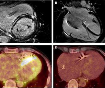
Cardiac sarcoidosis (CS), a rare condition characterized by non-caseating granulomas, can manifest with symptoms such as atrioventricular block and ventricular tachycardia (VT), as well as mimic inherited cardiomyopathies. A 58-year-old woman presented with sustained VT with a prior diagnosis of hypertrophic cardiomyopathy (HCM).
What is the utility of a head CT in cardiacarrest? We found intracranial hemorrhage in 2% of non-traumatic cardiacarrest patients, and in 4 others the presence of cerebral edema changed management. in Vienna found that 27 of 765 (4%) of out of hospital cardiacarrests (OHCA) were due to SAH.
High profile cases of sudden cardiacarrest in elite athletes in recent years has reminded the cardiology community of the challenging questions posed to cardiologists in these settings. Questions like: How do we prevent cardiacarrest in athletes? Can an athlete return to play after cardiacarrest?
And of course Ken's comments at the bottom) An elderly obese woman with cardiomyopathy, Left bundle branch block, and chronic hypercapnea presented hypoxic with altered mental status. Bedside cardiac ultrasound showed moderately decreased LV function. See this post: How a pause can cause cardiacarrest 2. The plan: 1.
Cardiac amyloidosis (CA) manifests as infiltrative cardiomyopathy and predisposes to sudden cardiacarrest (SCA), which can occur in about two-thirds of the patients with CA. An electrocardiogram (ECG) can be used as a readily available and affordable method that could assist in the risk stratification of the disease.
Cardiomyopathies: These diseases affect the heart muscle, impairing its ability to pump blood effectively. Mutations in specific genes often cause hypertrophic cardiomyopathy and dilated cardiomyopathy. How Do Genetic Factors Work?
It usually affects older patients suffering from coronary artery disease and cardiomyopathies. In cardiacarrest survivors, ventricular fibrillation (VF) is almost always the arrhythmic disorder identified at the time of the event.
CardiomyopathyCardiomyopathy is a condition that affects the heart muscle, causing it to become enlarged, thick, or rigid. Excessive Alcohol or Drug Use Long-term abuse of alcohol or certain drugs can weaken the heart muscle, resulting in cardiomyopathy and eventually cardiomegaly.
As per my review of this subject ( Check out My Comment at the bottom of the page in the November 16, 2023 post in Dr. Smith's ECG Blog ) — the 3 most common Causes of ACS ( A cute C oronary S yndrome ) with a "negative" cath are: i ) Myocarditis; ii ) Takotsubo cardiomyopathy; and , iii ) MINOCA.
Introduction:Stress Induced Cardiomyopathy is increasingly becoming more prevalent with increasing awareness about disease condition with annual incidence of 30 cases/100000 per year and an incidence of 1-2% in the patients presenting with acute coronary syndrome.[1] for stress induced cardiomyopathy and found 10450 patients in the data base.
The ECG in Figure-1 was obtained from an 18-year old woman — who moments before been resuscitated from out-of-hospital cardiacarrest. Does this ECG in Figure-1 provide clue(s) to the etiology of this patient's cardiacarrest? I suspected the answer resides in the reason why an 18-year woman might have a cardiacarrest.
Shortly after arrival in the ED ( E mergency D epartment ) — she suffered a cardiacarrest. BUT — Cardiac catheterization done a little later did not reveal any significant stenosis. Figure-1: The initial ECG in today's case — obtained after successful resuscitation from cardiacarrest. ( No CP ( C hest P ain ).
Although one may have all kinds of ischemic findings as a result of cardiacarrest (rather than cause of cardiacarrest), this degree of ST elevation and HATW is all but diagnostic of acute proximal LAD occlusion. This prompted cath lab activation. On arrival to the ED, this ECG was recorded: What do you think?
This list takes on new relevance given the ongoing Covid-19 pandemic — which predisposes to acute thrombotic events, stress cardiomyopathy ( Takotsubo ), infarction/ischemia and myocarditis. But a look at Figure-2 reminds us of a long list of additional entities to consider!
Background:Despite the role of cardiac catheterization for hemodynamic assessment and endomyocardial biopsy (EMB) in children with cardiomyopathy, data on procedure-related major adverse events (MAE) in this population is lacking. We aim to describe the rate of MAE in children with cardiomyopathy undergoing cardiac catheterization.
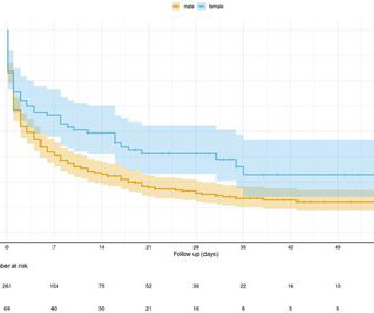
Introduction The use of venoarterial extracorporeal membrane oxygenation (VA-ECMO) in extracorporeal cardiopulmonary resuscitation (eCPR) has emerged as a treatment option for selected patients who are experiencing refractory cardiacarrest (CA). Results Out of the 377 patients included in the study, 69 (21%) were female.
Patients with hypertrophic cardiomyopathy (HCM) are enrolled in the national Cardiac Inherited Diseases Registry New Zealand. Thirteen probands (4%) presented with sudden death and 19 (6%) with cardiacarrest. Circulation: Heart Failure, Ahead of Print. BACKGROUND:Aotearoa/New Zealand has a multiethnic population.
METHODS:The AHA, through its Epidemiology and Prevention Statistics Committee, continuously monitors and evaluates sources of data on heart disease and stroke in the United States and globally to provide the most current information available in the annual Statistical Update with review of published literature through the year before writing.
Cardiac research scientist Dr. Sian Harding, on Takotsubo syndrome: "Natural disasters resulting in catastrophic stress almost always see a rise in both cardiacarrest and this mysterious heart condition."
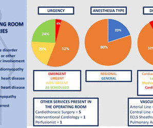
Patients were classified by CVD type: adult congenital heart disease, cardiacarrest, connective tissue disease with aortopathy, ischaemic cardiomyopathy, non-ischaemic cardiomyopathy or valve disease. We examined demographic, anaesthetic and procedure-related variables and in-hospital maternal and fetal outcomes.
Edited by Bracey, Meyers, Grauer, and Smith A 50-something-year-old female with a history of an unknown personality disorder and alcohol use disorder arrived via EMS following cardiacarrest with return of spontaneous circulation. The described rhythm was an irregular, wide complex rhythm.
And pretty much every doctor can recall an event where a patient experienced a suddenly stressful event and had a cardiac event. That event might have been a heart rhythm issue or even a cardiacarrest. In the hours after the 911 attacks on the World Trade Centre in New York, the rates of cardiacarrest more than doubled.
This phenomenon has been associated with cardiacarrest; after cardiac surgery; post-cardioversion following a sustained tachyarrhythmia; with certain types of acute cerebrovascular events ( such as subarachnoid hemorrhage ) — and , in association with large acute MI , which could potentially be the clinical scenario in today's case!
CardiacarrestCardiacarrest is a medical emergency in which the heart stops pumping blood to the body. Electrocardiogram, echocardiogram, and some other tests are done for patients with cardiacarrest. If the vital organs do not get their blood supply back quickly, it can lead to death.
Logistic regression was used to analyze the relationship between the treatment groups and hospital readmission within 90 days.Results:Only 517 AA met inclusion criteria and did not meet exclusion criteria, which included a history of valvular heart disease, hypertrophic or restrictive cardiomyopathy, active myocarditis, history of cardiacarrest, and (..)
Video-based AI A profound learning approach is created with a video-based neural system that utilizes a current database of video formats to determine cardiac issues. The deep learning algorithm helps segment the left ventricle predicting cardiomyopathy and ejection fraction. AI recognizing cardiacarrests in emergency calls.
CHD was the pre-PHT diagnosis in 21(62%) of stroke Pts, cardiomyopathy in 12(35%) and acquired heart disease in 1(3%). Data are presented as frequency (%) or median (Inter Quartile Range-IQR).Results:Of Median age of HT in these Pts was 6 yrs(1,13) (Table 1), 21(62%) were male.
It is apparently fortunate that she had a cardiacarrest; otherwise, her ECG would have been ignored. by making it clear to everyone that this is NOT an EKG that one sees with takotsubo cardiomyopathy. The impact of ST-segment elevation on the prognosis of patients with Takotsubo cardiomyopathy.
As discussed in ECG Blog #108 — AIVR generally occurs in one of the following C linical S ettings : i ) As a rhythm during cardiacarrest; ii ) In the monitoring phase of acute MI ( especially with inferior MI ) ; or , iii ) As a reperfusion arrhythmia ( ie, following thrombolysis, acute angioplasty, or spontaneous reperfusion ).
As always, takotsubo cardiomyopathy and focal pericarditis can mimic OMI, but takotsubo almost never mimics posterior MI, and both are diagnoses of exclusion after a negative cath. About two hours after admission, he suffered a cardiacarrest (whether it was VF/VT or PEA is not available) and expired.
A 50-something with severe chest pain and a normal ECG 30 Year Old with CardiacArrest, PEA, then Cardiac Ultrasound = Comment by K EN G RAUER, MD ( 12/18 /2022 ): = I thought the initial ECG in today's case ( which I've reproduced in Figure-1 ) — to be "eye-catching".
If a patient presents with chest pain and a normal heart rate, or with shockable cardiacarrest, then ischemic appearing ST elevation is STEMI until proven otherwise. Thus, there is a wall motion abnormality in the distribution of the LAD (not global apical dyskinesis, as in takostubo).
NOTE #3: In the context of a long QTc or ischemia — the finding of ST segment and/or T wave alternans may predict the occurrence of malignant ventricular arrhythmias.
The most important clinical entity associated with motion alternans is large pericardial effusion — though motion alternans has also been observed in some cases of hypertrophic cardiomyopathy. Conduction and Refractoriness Alternans — entails variance of impulse propagation along some par t of the conduction system.
We organize all of the trending information in your field so you don't have to. Join thousands of users and stay up to date on the latest articles your peers are reading.
You know about us, now we want to get to know you!
Let's personalize your content
Let's get even more personalized
We recognize your account from another site in our network, please click 'Send Email' below to continue with verifying your account and setting a password.


Let's personalize your content