This site uses cookies to improve your experience. To help us insure we adhere to various privacy regulations, please select your country/region of residence. If you do not select a country, we will assume you are from the United States. Select your Cookie Settings or view our Privacy Policy and Terms of Use.
Cookie Settings
Cookies and similar technologies are used on this website for proper function of the website, for tracking performance analytics and for marketing purposes. We and some of our third-party providers may use cookie data for various purposes. Please review the cookie settings below and choose your preference.
Used for the proper function of the website
Used for monitoring website traffic and interactions
Cookie Settings
Cookies and similar technologies are used on this website for proper function of the website, for tracking performance analytics and for marketing purposes. We and some of our third-party providers may use cookie data for various purposes. Please review the cookie settings below and choose your preference.
Strictly Necessary: Used for the proper function of the website
Performance/Analytics: Used for monitoring website traffic and interactions
Bedside cardiac ultrasound showed moderately decreased LV function. Discontinue all negative chronotropic agents, since the risk of torsade is much higher with bradycardia or pauses. She was intubated. CT of the chest showed no pulmonary embolism but bibasilar infiltrates. The plan: 1. Place temporary pacemaker 3.
Case continued Another ECG was recorded 3 hours later, still 1/10 pain: There is sinus bradycardia with RBBB. A bedside cardiac ultrasound performed by a true EM expert (Robert Reardon, who wrote the cardiac ultrasound chapter in Ma and Mateer) showed an inferior wall motion abnormality. They only mask the underlying pathology.
The ECG shows sinus bradycardia but is otherwise normal. On intravascular ultrasound (IVUS), the mid RCA plaque was described as "cratered, inflamed, and bulky," and the OM plaque was described as "bulky with evidence of inflammation and probably ulceration." The documentation does not describe any additional details of the history.
Here is his ED ECG: There is bradycardia with a junctional escape. Case continued A bedside ultrasound showed diminished LV EF and of course bradycardia. BP was 108 systolic (if a cuff pressure can be trusted) but appeared to be maintaining BP only by very high systemic vascular resistance. What is the atrial activity?
Use of drugs producing bradycardia like beta blockers in stages III and IV may precipitate low output state. J Cardiovasc Ultrasound. J Cardiovasc Ultrasound. In stage IV, this restrictive filling pattern remains fixed even during Valsalva maneuver. Initial stages (I to III) are considered reversible with treatment. Ha J et al.
Dr. Nossen performed a bedside ultrasound which was interpreted as normal. After the heart rate increased slightly, here was the repeat ECG: Sinus bradycardia, only slightly faster rate than prior. That said — what is unusual about the rhythm in the initial ECG of today's case — is the marked bradycardia!
There is sinus bradycardia with one PVC. The ways to tell for certain include intravascular ultrasound (to look for extra-luminal plaque with rupture) or "optical coherence tomography," something I am entirely unfamiliar with. She then had a 12-lead: What do you think?
Patient presentation is important This was a 60-something with acute chest pain: There is sinus bradycardia at a rate of 44. In case you were wondering about the T-waves and bradycardia, the K was normal. Why bradycardia? Maybe there is also inferior MI from wraparound LAD with associated sinus bradycardia.
Triage physician interpretation: -sinus bradycardia -lateral ST depressions While there are lateral ST depressions (V5, V6) the deepest ST depressions are in V4. The screening physician ordered an EKG and noted his ashen appearance and moderate distress. Triage EKG: What do you think? J Am Heart Assoc. 2021 Dec 7;10(23):e022866. 121.022866.
Her bedside cardiac ultrasound was normal We decided to cardiovert her since the time of onset was very recent. Baseline bradycardia in endurance athletes limits the use of ß-blockers. But when you see this, you should suspect that the AV node is not well. I signed her out to one of my partners.
Both of these features make inferior + RV MI by far the most likely ( Pseudoanteroseptal MI is another name for this ) There is also sinus bradycardia and t he patient is in shock with hypotension. A narrow complex bradycardia without any P-waves is also likely to respond to atropine, as it may be a junctional rhythm.
There was no evidence bradycardia leading up to the runs of PMVT ( as tends to occur with Torsades ). If there had been — a temporary atrial pacemaker could have been considered as a way of increasing the heart rate to suppress a bradycardia-dependent arrhythmia ("overdrive pacing").
Arrival at time 0 ECG 7 min Roomed in hallway at 17 min Moved to room with monitor at 37 min The patient was seen briefly by the physician, who then went to get an ultrasound machine. MY Thoughts on the ECG in Figure-1: The rhythm in ECG #1 is sinus bradycardia at ~50-55/minute. Then he was placed in a room after 30 minutes.
In any case, there is bradycardia. A bedside cardiac ultrasound was recorded: Here is a still image of the echo: The red arrows outline the right ventricle and the yellow arrows outline the left ventricle chamber. Second: what does the ultrasound tell us about the condition? No shock was ever delivered. He was in profound shock.
However the patient continued to have chest pain and bedside ultrasound showed hypokinesis of the septum with significantly reduced LVEF. About 2 hours later the patient arrived at a PCI-capable center and repeat ECG was obtained: The transferring EMS crew noted “runs of VT” during transport. Do not treat AIVR.
Bedside ultrasound showed no effusion and moderately decreased LV function, with B-lines of pulmonary edema. There is also bradycardia. Bradycardia puts patients at risk for "pause-dependent" Torsades de Pointes. Angio had shown some acute disease in the saphenous vein graft to the posterior descending artery off of the RCA.
Check : [vitals, SOB, Chest Pain, Ultrasound] If the patient has Abdominal Pain, Chest Pain, Dyspnea or Hypoxemia, Headache, Hypotension , then these should be considered the primary chief complaint (not syncope). Aortic Dissection, Valvular (especially Aortic Stenosis), Tamponade. Frequent or repetitive PACs ii. orthostatic vitals b.
Regardless of further evaluation, she should avoid bradycardia, AV nodal blockers, Na channel blockers, and fevers. --If A bedside cardiac ultrasound revealed grossly normal to hyperdynamic systolic function with no obvious areas of wall motion abnormalities.
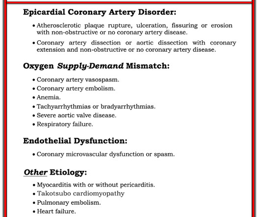
There is also STE in V1 which is diagnostic of right ventricular OMI in this situation , and partly explains the syncope and hypotension (along with the bradycardia). They recorded this ECG: Obvious inferior STEMI/OMI What else? The cath lab was activated by the medics. Etiologies (list not comprehensive): Coronary Spasm. Embolism with lysis.
Several days into hospitalization, she continued to have occasional episodes of sinus rhythm and sinus bradycardia with periods of Mobitz I AV block and 2:1 block. Meanwhile, the patient's native rhythm is sinus bradycardia with adequate perfusion. If you don't have ultrasound (but you should), then palpate a pulse!
We organize all of the trending information in your field so you don't have to. Join thousands of users and stay up to date on the latest articles your peers are reading.
You know about us, now we want to get to know you!
Let's personalize your content
Let's get even more personalized
We recognize your account from another site in our network, please click 'Send Email' below to continue with verifying your account and setting a password.


Let's personalize your content