This site uses cookies to improve your experience. To help us insure we adhere to various privacy regulations, please select your country/region of residence. If you do not select a country, we will assume you are from the United States. Select your Cookie Settings or view our Privacy Policy and Terms of Use.
Cookie Settings
Cookies and similar technologies are used on this website for proper function of the website, for tracking performance analytics and for marketing purposes. We and some of our third-party providers may use cookie data for various purposes. Please review the cookie settings below and choose your preference.
Used for the proper function of the website
Used for monitoring website traffic and interactions
Cookie Settings
Cookies and similar technologies are used on this website for proper function of the website, for tracking performance analytics and for marketing purposes. We and some of our third-party providers may use cookie data for various purposes. Please review the cookie settings below and choose your preference.
Strictly Necessary: Used for the proper function of the website
Performance/Analytics: Used for monitoring website traffic and interactions
EMS reports intermittent sinus tachycardia and bradycardia secondary to some type of heart block during transport. Limiting one's interpretation to marked bradycardia with high-grade AV block in need of pacing in this patient with multiple syncopal epiosodes — more than suffices for "the quick answer".
In usual syncope, there could be a fall in blood pressure, bradycardia, and there are various types, which will be described. The most common is mixed type, in which, in the tilted position, the person develops both bradycardia and hypotension and usually there is a syncope. Various types of responses can occur to head up tilt test.
They had dense calcifications at the lesions, stenosis rates of 95% (near occlusion) and 86% according to the North American Symptomatic Carotid Endarterectomy Trial criteria, and calcification arcs of 270° and 360°, respectively. After postdilatation, the stenosis rates decreased to 21% and 23%, respectively.
Angiography revealed a 30% nonobstructive stenosis of the mid LAD. He had multiple episodes of bradycardia and nonsustained ventricular tachycardia. There was a 70% culprit stenosis of the first obtuse marginal branch in a right dominant system. Serial high sensitivity troponin T (URL 15 ng/L) values were negative and stagnant.
The computer called "Sinus Bradycardia" only (implying that everything else is normal. The overreading Cardiologist called it only "Sinus Bradycardia" with no other findings. The rhythm in Figure-1 is sinus bradycardia and arrhythmia. Here is the old ECG from 6 years prior: Notice the inferior T-waves have normal size here.
The ECG shows sinus bradycardia but is otherwise normal. The LAD has diffuse disease with a few areas of moderate stenosis but no flow-limiting lesions. Written by Willy Frick A 46 year old man with a history of type 2 diabetes mellitus presented to urgent care with complaint of "chest burning." The following ECG was obtained.
Angiogram showed a critical LAD thrombotic stenosis. The patient went to cath and had a distal LAD 99% stenosis with thrombus and TIMI-2 flow. Patient presentation is important This was a 60-something with acute chest pain: There is sinus bradycardia at a rate of 44. Why bradycardia? He underwent CABG. Peak was 8.1
The cath lab was activated: Result: Thrombotic 95% stenosis at the ostium of a small LPL2 with 70% stenosis at the LPL2/LPDA bifurcation in the distal/AV groove Cx Tubular 70% stenosis in the mid-circumflex. (In This is sinus bradycardia. There was concern for aortic dissection, so a CT was done and was negative.
Here is his previous ECG: This was my interpretation of the first ECG: Sinus bradycardia with less than 1mm ST elevation in V4-V6, elevated compared to the previous ECG, suggestive of lateral MI. There is evidence that de Winter's T-waves really represent a tiny trickle of blood through the thrombotic stenosis.
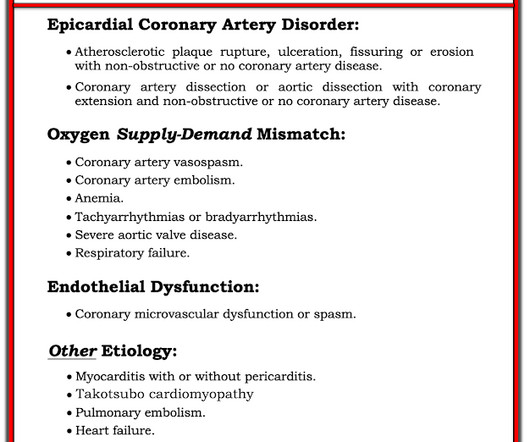
There is sinus bradycardia with one PVC. distal stenosis or occluded small branches), and 3) nonischemic causes for myocyte injury (e.g., She then had a 12-lead: What do you think? The diagnosis of MINOCA should exclude: 1) other overt causes for elevated troponin (e.g., pulmonary embolism, sepsis, etc.), myocarditis).
Triage physician interpretation: -sinus bradycardia -lateral ST depressions While there are lateral ST depressions (V5, V6) the deepest ST depressions are in V4. 90% stenosis of the proximal ramus intermedius, pre procedure TIMI II flow The ramus intermedius is a normal variant on coronary anatomy that arises between the LAD and LCX.
found that such ECG findings only represented left main ACS in 14% of such ECGs: Only 23% of patients with the aVR STE pattern had any LM disease (fewer if defined as 50% stenosis). In my experience, Ive seen U waves not only with low K+/low Mg++ but also in patients with bradycardia, LVH, and sometimes in normal subjects.
I said I think there is a fixed stenosis in the LAD and the tachycardia and stress caused a type 2 STEMI. In this abstract from 2011, we found that 4%(4 of 99) type 2 MI and 38% of type 1 MI had ST Elevation. link] An angiogram was done: It showed no culprit and no coronary disease, but did show a myocardial bridge in the mid LAD.
Both of these features make inferior + RV MI by far the most likely ( Pseudoanteroseptal MI is another name for this ) There is also sinus bradycardia and t he patient is in shock with hypotension. A narrow complex bradycardia without any P-waves is also likely to respond to atropine, as it may be a junctional rhythm.
Angiography : LMCA — 90-99% osteal stenosis. LCx — 50-69% stenosis of the 1st marginal branch; with 100% distal LCx occlusion. There was no evidence bradycardia leading up to the runs of PMVT ( as tends to occur with Torsades ). The image shows the impella device in place. RCA — 100% proximal occlussion.
Below is a still image with the red arrow indicating the subtotal LMCA stenosis. As per Dr. Nossen — today's initial ECG ( LEFT tracing in Figure-2 ) shows sinus bradycardia with QRS widening due to bifascicular block ( RBBB/LAHB ). The video below shows the coronary angiography. Pressors could gradually be tapered within 24 hours.
Reasons for not prescibing or discontinuing were: CKD 6, severe aortic stenosis 5, asthma 3, symptomatic bradycardia 5, hypotension 3, type1 diabetes 2, syncope 1, Raynauds 1, patient choice 8 and 6 patients died before all appropriate medications could be initiated. In 10 cases no clinical reason could be identified.It
Soon afterward, the patient’s symptoms return along with lightheadedness, bradycardia, and hypotension. The patient has also developed sinus bradycardia, which may result from right coronary artery ischemia to the SA node. The Queen of Hearts agrees: Around this time his initial high sensitivity troponin I resulted at 231 ng/L.
Aortic Dissection, Valvular (especially Aortic Stenosis), Tamponade. PVCs N ot generally considered abnormal ECG findings: Isolated PAC, First Degree AV Block, Sinus bradycardia at a rate of 35-45, and Nonspecific ST-T abnormalities (even if different from a previous ECG). heart auscultation (aortic stenosis); c.
There is also STE in V1 which is diagnostic of right ventricular OMI in this situation , and partly explains the syncope and hypotension (along with the bradycardia). They recorded this ECG: Obvious inferior STEMI/OMI What else? The cath lab was activated by the medics. As I wrote in that Nov.
We organize all of the trending information in your field so you don't have to. Join thousands of users and stay up to date on the latest articles your peers are reading.
You know about us, now we want to get to know you!
Let's personalize your content
Let's get even more personalized
We recognize your account from another site in our network, please click 'Send Email' below to continue with verifying your account and setting a password.


Let's personalize your content