This site uses cookies to improve your experience. To help us insure we adhere to various privacy regulations, please select your country/region of residence. If you do not select a country, we will assume you are from the United States. Select your Cookie Settings or view our Privacy Policy and Terms of Use.
Cookie Settings
Cookies and similar technologies are used on this website for proper function of the website, for tracking performance analytics and for marketing purposes. We and some of our third-party providers may use cookie data for various purposes. Please review the cookie settings below and choose your preference.
Used for the proper function of the website
Used for monitoring website traffic and interactions
Cookie Settings
Cookies and similar technologies are used on this website for proper function of the website, for tracking performance analytics and for marketing purposes. We and some of our third-party providers may use cookie data for various purposes. Please review the cookie settings below and choose your preference.
Strictly Necessary: Used for the proper function of the website
Performance/Analytics: Used for monitoring website traffic and interactions
CT of the chest showed no pulmonary embolism but bibasilar infiltrates. Discontinue all negative chronotropic agents, since the risk of torsade is much higher with bradycardia or pauses. Place temporary pacemaker 3. She was intubated. Bedside cardiac ultrasound showed moderately decreased LV function. The plan: 1.
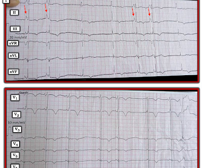
This middle-aged patient presented with SOB, weakness, and mild pulmonary edema. There are 3 etiologies I always think of with bradycardia and AV block: 1. She had a permanent pacemaker implanted. She previously had Atrial fibrillation with LBBB. Here is her ED ECG: Does this reveal the etiology of her symptoms? Hyperkalemia.
I’d guess the overall rhythm is sinus, perhaps with a wandering atrial pacemaker and very frequent ventricular ectopy with multiple couplets. Perhaps the patient has pulmonary hypertension and/or tricuspid regurgitation? In the meantime, a pacemaker may be needed. =
This is demonstrated ( Figure 5 ) by the gap in arrows at the bottom of the strip, signifying that the demand pacemaker has recognized an underlying rhythm (in this case, artifact from a moving ambulance). The artifact fools the pacemaker into thinking the rhythm is native.
PVCs N ot generally considered abnormal ECG findings: Isolated PAC, First Degree AV Block, Sinus bradycardia at a rate of 35-45, and Nonspecific ST-T abnormalities (even if different from a previous ECG). Thus, if there is documented sinus bradycardia, and no suspicion of high grade AV block, at the time of the syncope, this is very useful.
We organize all of the trending information in your field so you don't have to. Join thousands of users and stay up to date on the latest articles your peers are reading.
You know about us, now we want to get to know you!
Let's personalize your content
Let's get even more personalized
We recognize your account from another site in our network, please click 'Send Email' below to continue with verifying your account and setting a password.


Let's personalize your content