This site uses cookies to improve your experience. To help us insure we adhere to various privacy regulations, please select your country/region of residence. If you do not select a country, we will assume you are from the United States. Select your Cookie Settings or view our Privacy Policy and Terms of Use.
Cookie Settings
Cookies and similar technologies are used on this website for proper function of the website, for tracking performance analytics and for marketing purposes. We and some of our third-party providers may use cookie data for various purposes. Please review the cookie settings below and choose your preference.
Used for the proper function of the website
Used for monitoring website traffic and interactions
Cookie Settings
Cookies and similar technologies are used on this website for proper function of the website, for tracking performance analytics and for marketing purposes. We and some of our third-party providers may use cookie data for various purposes. Please review the cookie settings below and choose your preference.
Strictly Necessary: Used for the proper function of the website
Performance/Analytics: Used for monitoring website traffic and interactions
No ischemia. Case continued Another ECG was recorded 3 hours later, still 1/10 pain: There is sinus bradycardia with RBBB. This is a conundrum, because it is clear that the patient is having an acute MI, the ECG is dynamic, but the pain is very mild and there is no ECG evidence of active transmural ischemia.
Monomorphic ventricular tachycardia in the setting of acute myocardialischemia can also be treated by intravenous lignocaine bolus followed by infusion. Predisposing causes for ventricular tachycardia like ischemia and electrolyte imbalance has to be treated simultaneously to prevent recurrence.
Sinus bradycardia, normal conduction, normal axis, normal R wave progression, no hypertrophy. 2] Here there is no posterior ST elevation, but the anterior ST depression is also less—so it is dynamic, confirming acute ischemia. What do you think? But it is still STEMI negative.
WPW, previous Q wave MI, and acute coronary occlusion Depending on the location of the accessory pathway, WPW pattern can mimic ventricular hypertrophy (including RVH or LVH) or myocardialinfarction (including anterior, inferior, lateral or posterior MI) [1]. Wolff-Parkinson-White syndrome ‘cured’ by myocardialinfarction?
Here are inferior leads, and aVL, magnified: A closer inspection of the inferior leads and aVL Sinus bradycardia. I had no history on the case and no prior ECG for comparison. What do you think? The T-wave in lead III is slightly tall and broad (increased area under the curve) compared to its QRS complex.
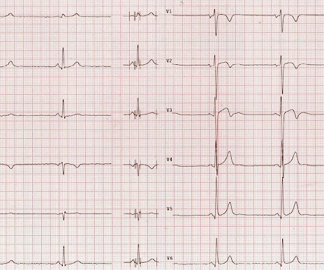
Here is his ECG: There is no clear evidence of OMI or ischemia. The Initial ECG in Today's Case: ECG #1 showed sinus bradycardia at a rate slightly under 60/minute — normal intervals — slight left axis ( about -15 degrees ) — and no chamber enlargement. A 40-something male with no previous cardiac disease presented with chest pain.
Electrocardiographic Differentiation of Early Repolarization FromSubtle Anterior ST-Segment Elevation MyocardialInfarction. Patient presentation is important This was a 60-something with acute chest pain: There is sinus bradycardia at a rate of 44. In case you were wondering about the T-waves and bradycardia, the K was normal.
Triage physician interpretation: -sinus bradycardia -lateral ST depressions While there are lateral ST depressions (V5, V6) the deepest ST depressions are in V4. Ischemic ST-Segment Depression Maximal in V1-V4 (Versus V5-V6) of Any Amplitude Is Specific for Occlusion MyocardialInfarction (Versus Nonocclusive Ischemia).
Both of these features make inferior + RV MI by far the most likely ( Pseudoanteroseptal MI is another name for this ) There is also sinus bradycardia and t he patient is in shock with hypotension. A narrow complex bradycardia without any P-waves is also likely to respond to atropine, as it may be a junctional rhythm.
AIVR is not always the result of significant pathology, but is classically associated with the reperfusion phase of acute myocardialinfarction. Similarly, the OMI paradigm respects ACS as a dynamic process in which ECG changes reflect the phase of myocardial injury and risk stratify which patients may benefit from emergent PCI.
There is also bradycardia. Bradycardia puts patients at risk for "pause-dependent" Torsades de Pointes. Torsades in acquired long QT is much more likely in bradycardia because the QT interval following a long pause is longer still. Literature on Hypokalemia as a risk for ventricular fibrillation in acute myocardialinfarction.
Troponin T peaked at 38,398 ng/L ( = a very large myocardialinfarction, but not massive-- thanks to the pre-PCI spontaneous reperfusion, and rapid internvention!! ). There is no definite evidence of acute ischemia. (ie, Some residual ischemia in the infarct border might still be present.
Description Sinus bradycardia. There is ST elevation in V2 and V3 There are inverted T-waves in V2 and V3 There are prominent U-waves in V2 and V3 Many responders were worried about ischemia or hypertrophic cardiomyopathy. This short QT at least makes ischemia all but impossible. There is high voltage. This is a normal variant.
Such findings would normally suggest primary ischemia with concomitant surveillance of coronary occlusion, but these ST/T changes might very well be secondary to the Escape mechanism at hand. Comparison of the QRS complex, ST-segment, and T-wave among patients with left bundle branch block with and without acute myocardialinfarction.
There’s sinus bradycardia, first degree AV block, normal axis, delayed R wave progression, and normal voltages. Hyperacute T waves are deflating, suggesting reperfusion but there is still reciprocal change in I/aVL and ST depression in V2, and the bradycardia is worse. Below is the ECG. What do you think? Take home 1. second ).
Followup ECG: No Change Absence of evolution is the best evidence against ischemia as the etiology. I was taught that the tell-tale sign of ischemia vs an electrical abnormality was in the hx, i.e. chest pain for the ischemia and potential syncope for brugada. Ischemia/infarction. Bradycardia. Hypothermia.
Evidence of acute ischemia (may be subtle) vii. PVCs N ot generally considered abnormal ECG findings: Isolated PAC, First Degree AV Block, Sinus bradycardia at a rate of 35-45, and Nonspecific ST-T abnormalities (even if different from a previous ECG). Old myocardialinfarction, 6. Left BBB vi. Pathologic Q-waves viii.
Despite the baseline artifact theres sinus bradycardia, convex ST elevation in III, reciprocal ST depression in aVL and possible anterior ST depression indicating inferoposterior OMI. Prevalence and outcome of patients with non-ST segment elevation myocardialinfarction with occluded culprit artery - a systemic review and meta-analysis.
This ECG shows a sinus bradycardia with a normal conduction pattern (normal PR, normal QRS, and normal QTc), normal axis, normal R-wave progression, normal voltages. Clinical questions : Is this an occlusion myocardialinfarction and does the patient need the cath lab? What do you think? Pacing Clin Electrophysiol.
Within ten minutes, she developed bradycardia, hypotension, and ST changes on monitor. If this were OMI, I would favor proximal RCA culprit (since that commonly produces inferolateral changes and occasionally produces anterior HATW from RV infarct ), but LAD is also possible. Bradycardia and heart block are very common in RCA OMI.
We organize all of the trending information in your field so you don't have to. Join thousands of users and stay up to date on the latest articles your peers are reading.
You know about us, now we want to get to know you!
Let's personalize your content
Let's get even more personalized
We recognize your account from another site in our network, please click 'Send Email' below to continue with verifying your account and setting a password.


Let's personalize your content