This site uses cookies to improve your experience. To help us insure we adhere to various privacy regulations, please select your country/region of residence. If you do not select a country, we will assume you are from the United States. Select your Cookie Settings or view our Privacy Policy and Terms of Use.
Cookie Settings
Cookies and similar technologies are used on this website for proper function of the website, for tracking performance analytics and for marketing purposes. We and some of our third-party providers may use cookie data for various purposes. Please review the cookie settings below and choose your preference.
Used for the proper function of the website
Used for monitoring website traffic and interactions
Cookie Settings
Cookies and similar technologies are used on this website for proper function of the website, for tracking performance analytics and for marketing purposes. We and some of our third-party providers may use cookie data for various purposes. Please review the cookie settings below and choose your preference.
Strictly Necessary: Used for the proper function of the website
Performance/Analytics: Used for monitoring website traffic and interactions
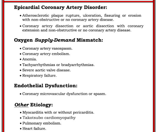
There is sinus bradycardia with one PVC. MINOCA: MyocardialInfarction in the Absence of Obstructive Coronary Artery Disease). pulmonary embolism, sepsis, etc.), Coronary thrombosis or embolism can result in MINOCA, either with or without a hypercoagulable state. She then had a 12-lead: What do you think?
Smith , d and Muzaffer Değertekin a DIFOCCULT: DIagnostic accuracy oF electrocardiogram for acute coronary OCClUsion resuLTing in myocardialinfarction. His first electrocardiogram ( ECG) is given below: --Sinus bradycardia. Bi-phasic scan showed no dissection or pulmonary embolism. References 1. Turk Kardiyol Dern Ars.
PVCs N ot generally considered abnormal ECG findings: Isolated PAC, First Degree AV Block, Sinus bradycardia at a rate of 35-45, and Nonspecific ST-T abnormalities (even if different from a previous ECG). Thus, if there is documented sinus bradycardia, and no suspicion of high grade AV block, at the time of the syncope, this is very useful.
There is also STE in V1 which is diagnostic of right ventricular OMI in this situation , and partly explains the syncope and hypotension (along with the bradycardia). Here it is annotated in red: Our extremely smart radiologist, Gopal Punjabi , assures me that this finding can only be due to myocardialinfarction, not myocarditis.
Within ten minutes, she developed bradycardia, hypotension, and ST changes on monitor. If this were OMI, I would favor proximal RCA culprit (since that commonly produces inferolateral changes and occasionally produces anterior HATW from RV infarct ), but LAD is also possible. Bradycardia and heart block are very common in RCA OMI.
We organize all of the trending information in your field so you don't have to. Join thousands of users and stay up to date on the latest articles your peers are reading.
You know about us, now we want to get to know you!
Let's personalize your content
Let's get even more personalized
We recognize your account from another site in our network, please click 'Send Email' below to continue with verifying your account and setting a password.


Let's personalize your content