This site uses cookies to improve your experience. To help us insure we adhere to various privacy regulations, please select your country/region of residence. If you do not select a country, we will assume you are from the United States. Select your Cookie Settings or view our Privacy Policy and Terms of Use.
Cookie Settings
Cookies and similar technologies are used on this website for proper function of the website, for tracking performance analytics and for marketing purposes. We and some of our third-party providers may use cookie data for various purposes. Please review the cookie settings below and choose your preference.
Used for the proper function of the website
Used for monitoring website traffic and interactions
Cookie Settings
Cookies and similar technologies are used on this website for proper function of the website, for tracking performance analytics and for marketing purposes. We and some of our third-party providers may use cookie data for various purposes. Please review the cookie settings below and choose your preference.
Strictly Necessary: Used for the proper function of the website
Performance/Analytics: Used for monitoring website traffic and interactions
CT of the chest showed no pulmonary embolism but bibasilar infiltrates. Discontinue all negative chronotropic agents, since the risk of torsade is much higher with bradycardia or pauses. She was intubated. Bedside cardiac ultrasound showed moderately decreased LV function. The plan: 1. Place temporary pacemaker 3.
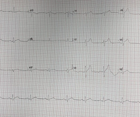
This ECG was recorded on arrival in the ED: Here is the interpretation of the conventional algorithm (Veritas): SINUS BRADYCARDIA ST ELEVATION, PROBABLY EARLY REPOLARIZATION [ST ELEVATION WITH NORMALLY INFLECTED T-WAVE] BORDERLINE ECG What do you think?
The receiving staff suspects pulmonary embolism due to S1Q3T3 on the ECG and administers TPA. Learning points: TCP is primarily recommended for bradycardia that does not respond to atropine, or other agents. The patient did have massive pulmonary emboli, but he also had profound intraventricular and subarachnoid hemorrhages.
In any case, there is bradycardia. It makes pulmonary embolism (PE) very likely. But it is bradyasystolic, so pulmonary embolism must be high on the differential. Possible, but huge pulmonary embolism is more likely. Such isolated RV STEMI is rare, but pulmonary embolism is not. No shock was ever delivered.
Normal 0 false false false EN-US X-NONE X-NONE Normal 0 false false false EN-US X-NONE X-NONE The final diagnosis on his ED note: pulmonary embolism AND pericarditis. Does subsegmental pulmonary embolism matter? The ST/T ratio in V6, however, is slightly greater. How do we know that the benefits outweigh the risks of anticoagulation?
His first electrocardiogram ( ECG) is given below: --Sinus bradycardia. Bi-phasic scan showed no dissection or pulmonary embolism. Blood pressure: 130/80 mmHg, heart rate: 45/min, respiratory rate: 18/min, SaO2: %98, body temperature: normal. As he seemed very agitated, fentanyl and diazepam were given.
There is sinus bradycardia with one PVC. pulmonary embolism, sepsis, etc.), Coronary thrombosis or embolism can result in MINOCA, either with or without a hypercoagulable state. She then had a 12-lead: What do you think? The diagnosis of MINOCA should exclude: 1) other overt causes for elevated troponin (e.g., myocarditis).
PVCs N ot generally considered abnormal ECG findings: Isolated PAC, First Degree AV Block, Sinus bradycardia at a rate of 35-45, and Nonspecific ST-T abnormalities (even if different from a previous ECG). Thus, if there is documented sinus bradycardia, and no suspicion of high grade AV block, at the time of the syncope, this is very useful.
Within ten minutes, she developed bradycardia, hypotension, and ST changes on monitor. Bradycardia and heart block are very common in RCA OMI. Third, a slow motion segment showing delayed, brisk filling of the PDA due to dislodgment of a thrombus from contrast injection and distal embolization.
We organize all of the trending information in your field so you don't have to. Join thousands of users and stay up to date on the latest articles your peers are reading.
You know about us, now we want to get to know you!
Let's personalize your content
Let's get even more personalized
We recognize your account from another site in our network, please click 'Send Email' below to continue with verifying your account and setting a password.


Let's personalize your content