This site uses cookies to improve your experience. To help us insure we adhere to various privacy regulations, please select your country/region of residence. If you do not select a country, we will assume you are from the United States. Select your Cookie Settings or view our Privacy Policy and Terms of Use.
Cookie Settings
Cookies and similar technologies are used on this website for proper function of the website, for tracking performance analytics and for marketing purposes. We and some of our third-party providers may use cookie data for various purposes. Please review the cookie settings below and choose your preference.
Used for the proper function of the website
Used for monitoring website traffic and interactions
Cookie Settings
Cookies and similar technologies are used on this website for proper function of the website, for tracking performance analytics and for marketing purposes. We and some of our third-party providers may use cookie data for various purposes. Please review the cookie settings below and choose your preference.
Strictly Necessary: Used for the proper function of the website
Performance/Analytics: Used for monitoring website traffic and interactions
Electrocardiogram (ECG) and telemetry revealed junctional bradycardia with heart rate in 30s and sinus pauses (5-7 seconds). He was admitted for further workup of bradycardia. His home medications included metoprolol succinate 25mg daily which was held given bradycardia. Echocardiogram was unchanged from baseline.
Discontinue all negative chronotropic agents, since the risk of torsade is much higher with bradycardia or pauses. EKG with paced complexes shown below shows much narrower QRS complex and echocardiogram showed improved LV systolic function primarily due to improvement in LV dyssynchrony. (J The plan: 1. Place temporary pacemaker 3.
EMS reports intermittent sinus tachycardia and bradycardia secondary to some type of heart block during transport. The echocardiogram showed a normal EF without any abnormalities. Each event is associated with a prodrome of mild substernal CP, SOB, and “brain fog.” Troponins were all negative.
Hopefully a repeat echocardiogram will be performed outpatient. Other Arrhythmias ( PACs, PVCs, AFib, Bradycardia and AV conduction disorders — potentially lethal VT/VFib ). Systolic function normal by visual assessment only, unable to visualize well for further characterization. 1900: RBBB and LAFB are almost fully resolved.
Additionally, a bedside echocardiogram showed no wall motion abnormality and normal LV function. He had multiple episodes of bradycardia and nonsustained ventricular tachycardia. A formal echocardiogram for patient 2 showed normal LV size, wall thickness, and global systolic function.
Patients who received pacemakers for an advanced atrioventricular block or bradycardia with atrial fibrillation, baseline LV ejection fraction (LVEF) ≥ 50%, and echocardiogram recorded at least 6 months postimplantation were included. The paced QRS recorded immediately after implantation was analyzed.
The diagnosis was a bit hard to find in the chart, and the echocardiogram did only stated "assymetric hypertrophy." It turns out that she has hypertrophic cardiomyopathy.
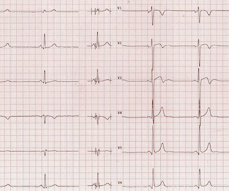
The computer called "Sinus Bradycardia" only (implying that everything else is normal. The overreading Cardiologist called it only "Sinus Bradycardia" with no other findings. Here is the post PCI EKG: And a few hours after that: The post PCI echocardiogram showed: Normal estimated left ventricular ejection fraction, 57%.
Due to limitations of echocardiogram in evaluating the right ventricle, magnetic resonance imaging study of the right ventricle along with that of the left ventricle has been reported. Athlete’s bradycardia due to increased parasympathetic tone and decreased sympathetic tone is a well-known observation.
5 years ago Similar Previous formal echocardiogram Inferior posterior with dyskinesis "Dyskinesis" is the technical echo term for LV aneurysm. Over time, T-waves normalize in the absence of new OMI. So upright T-waves in the presentation ECG do NOT mean there is any re-occlusion.
An echocardiogram was done. Other Arrhythmias ( PACs, PVCs, AFib, Bradycardia and AV conduction disorders — potentially lethal VT/VFib ). Is there also Brugada? Here is the result: The estimated left ventricular ejection fraction is 50 %. There is no left ventricular wall motion abnormality identified. Right ventricular prominence.
The relationship between low RHR and CI has yet to be described.Purpose:We hypothesize that resting sinus bradycardia (low RHR) could be a predictor of chronotropic incompetence and reduced exercise capacity.Methods:The derivation cohort consists of 201 patients with normal Bruce protocol treadmill stress echocardiogram.
Description Sinus bradycardia. First because I have a good eye on ECGs of endurance athletes Second because I see a lot of these tracings Third because the stress test determines the disappearance of ECG abnor malities found at rest Fourth because the echocardiogram is normal Fifth and last, the clinical presentation speaks clearly."
However, an echocardiogram is a different test, also conducted for heart activity. A fast heartbeat is called tachycardia, while a slow heartbeat is called bradycardia in medical terms. Electrocardiogram, echocardiogram, and some other tests are done for patients with cardiac arrest. ECG and EKG refer to the same thing.
He visited an outpatient clinic for it and an echocardiogram and exercise stress test was normal. His first electrocardiogram ( ECG) is given below: --Sinus bradycardia. In the meantime, cardiology consultant sees the patient and performs a bedside echocardiogram which revealed no major wall motion abnormalities.
Soon afterward, the patient’s symptoms return along with lightheadedness, bradycardia, and hypotension. The patient has also developed sinus bradycardia, which may result from right coronary artery ischemia to the SA node. The Queen of Hearts agrees: Around this time his initial high sensitivity troponin I resulted at 231 ng/L.
A formal echocardiogram was completed the next day and again showed a normal ejection fraction without any focal wall motion abnormalities to suggest CAD. Cardiology was consulted and they agreed that the EKG had an atypical morphology for STEMI and did not activate the cath lab.
PVCs N ot generally considered abnormal ECG findings: Isolated PAC, First Degree AV Block, Sinus bradycardia at a rate of 35-45, and Nonspecific ST-T abnormalities (even if different from a previous ECG). Thus, if there is documented sinus bradycardia, and no suspicion of high grade AV block, at the time of the syncope, this is very useful.
There are 2 main options: Overdrive pacing could be considered and in the right clinical situation, this is often effective for reducing ventricular arrhythmias ( especially in the case of preventing pause induced or bradycardia-induced arrhythmias in association with QTc prolongation ). Try a different kind of antiarrhythmic.
We organize all of the trending information in your field so you don't have to. Join thousands of users and stay up to date on the latest articles your peers are reading.
You know about us, now we want to get to know you!
Let's personalize your content
Let's get even more personalized
We recognize your account from another site in our network, please click 'Send Email' below to continue with verifying your account and setting a password.


Let's personalize your content