This site uses cookies to improve your experience. To help us insure we adhere to various privacy regulations, please select your country/region of residence. If you do not select a country, we will assume you are from the United States. Select your Cookie Settings or view our Privacy Policy and Terms of Use.
Cookie Settings
Cookies and similar technologies are used on this website for proper function of the website, for tracking performance analytics and for marketing purposes. We and some of our third-party providers may use cookie data for various purposes. Please review the cookie settings below and choose your preference.
Used for the proper function of the website
Used for monitoring website traffic and interactions
Cookie Settings
Cookies and similar technologies are used on this website for proper function of the website, for tracking performance analytics and for marketing purposes. We and some of our third-party providers may use cookie data for various purposes. Please review the cookie settings below and choose your preference.
Strictly Necessary: Used for the proper function of the website
Performance/Analytics: Used for monitoring website traffic and interactions
Electrocardiogram (ECG) and telemetry revealed junctional bradycardia with heart rate in 30s and sinus pauses (5-7 seconds). He was admitted for further workup of bradycardia. His home medications included metoprolol succinate 25mg daily which was held given bradycardia. Initial laboratory analysis was unremarkable.
We talk about the ketogenic diet as a metabolic therapy for type 1 diabetes. Dozens if not hundreds of (albeit observational) studies have linked a short sleep duration to a number of health conditions including type 2 diabetes and cardiovascular disease. Welcome to the Physiology Friday newsletter.
Written by Willy Frick A 46 year old man with a history of type 2 diabetes mellitus presented to urgent care with complaint of "chest burning." The ECG shows sinus bradycardia but is otherwise normal. The documentation does not describe any additional details of the history. The following ECG was obtained. ECG 1 What do you think?
He denied any known medical history, specifically: coronary artery disease, hypertension, dyslipidemia, diabetes, heart failure, myocardial infarction, or any prior PCI/stent. Breath sounds were clear in all lung fields. No appreciable skin pallor. He reported to be a social drinker, but used tobacco products daily.
He has a history of known CAD, diabetes, and dyslipidemia. Here is his previous ECG: This was my interpretation of the first ECG: Sinus bradycardia with less than 1mm ST elevation in V4-V6, elevated compared to the previous ECG, suggestive of lateral MI. By pure clinical appearance, he looked like the textbook patient with acute MI.
A 50-something male with unspecified history of cardiomyopathy presented in diabetic ketoacidosis (without significant hyperkalemia) with a wide complex tachycardia and hypotension. The patient later settled into sinus bradycardia. Bedside echo showed "mildly reduced" LV EF. Here is the ED ECG: What do you think? It is regular.
Biphasic T-waves in a Middle-Aged Male with Vomiting Diabetic Ketoacidosis: is there hypokalemia? In my experience, Ive seen U waves not only with low K+/low Mg++ but also in patients with bradycardia, LVH, and sometimes in normal subjects. You probably think it is left main. Are These Wellens' Waves?? ST depression: is it ischemia?
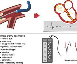
Background Rotational atherectomy (RA) during percutaneous coronary intervention may cause transient bradycardia or a higher-degree heart block. Traditionally, some operators use prophylactic transvenous pacing wire (TPW) to avoid haemodynamic complications associated with bradycardia. No patients underwent prophylactic TPW insertion.
Our collaboration with Orchestra BioMed will explore how cardiac pacing can go beyond management of bradycardia and conduction disease to treat hypertension as well,” said Robert C. Kowal, M.D., vice president and general manager of Cardiac Pacing Therapies within the Medtronic Cardiac Rhythm Management operating unit.
Written by Magnus Nossen The patient in today's case is a male in his 70s with hypertension and type II diabetes mellitus. As per Dr. Nossen — today's initial ECG ( LEFT tracing in Figure-2 ) shows sinus bradycardia with QRS widening due to bifascicular block ( RBBB/LAHB ). The syncope lasted about 2-3 minutes according to his wife.
Reasons for not prescibing or discontinuing were: CKD 6, severe aortic stenosis 5, asthma 3, symptomatic bradycardia 5, hypotension 3, type1 diabetes 2, syncope 1, Raynauds 1, patient choice 8 and 6 patients died before all appropriate medications could be initiated. In 10 cases no clinical reason could be identified.It
In isolation, however, syncope does not hold significant weight for OMI – as opposed to something like crushing chest discomfort, for example – although stereotypical ACS might become blurry in both the elderly and diabetic populations. This is important because we must rely on the ECG to further elucidate the story when the patient cannot.
Written by Jesse McLaren An 80 year old patient with diabetes/hypertension/ cirrhosis had a recent increase in candesartan for their hypertension, and was also on spirolactone and nadolol. Theres no prior ECG to compare - but the bradycardia, prolonged PR and peaked T waves could all be from hyperkalemia. Take away 1.
We organize all of the trending information in your field so you don't have to. Join thousands of users and stay up to date on the latest articles your peers are reading.
You know about us, now we want to get to know you!
Let's personalize your content
Let's get even more personalized
We recognize your account from another site in our network, please click 'Send Email' below to continue with verifying your account and setting a password.


Let's personalize your content